• 文献检索
  • 文档翻译
  • 深度研究
  • 学术资讯
  • Suppr Zotero 插件Zotero 插件
  • 邀请有礼
  • 套餐&价格
  • 历史记录
应用&插件
Suppr Zotero 插件Zotero 插件浏览器插件Mac 客户端Windows 客户端微信小程序
定价
高级版会员购买积分包购买API积分包
服务
文献检索文档翻译深度研究API 文档MCP 服务
关于我们
关于 Suppr公司介绍联系我们用户协议隐私条款
关注我们

Suppr 超能文献

核心技术专利:CN118964589B侵权必究
粤ICP备2023148730 号-1Suppr @ 2026

文献检索

告别复杂PubMed语法,用中文像聊天一样搜索,搜遍4000万医学文献。AI智能推荐,让科研检索更轻松。

立即免费搜索

文件翻译

保留排版,准确专业,支持PDF/Word/PPT等文件格式,支持 12+语言互译。

免费翻译文档

深度研究

AI帮你快速写综述,25分钟生成高质量综述,智能提取关键信息,辅助科研写作。

立即免费体验

骨桥蛋白水平与多发性硬化症晚期区域性脑容量降低有关。

Osteopontin levels are associated with late-time lower regional brain volumes in multiple sclerosis.

机构信息

MTA-PTE Clinical Neuroscience MR Research Group, Eötvös Loránd Research Network (ELKH), Ret str. 2, 7623, Pecs, Hungary.

Department of Neurology, Medical School, University of Pecs, Pecs, Hungary.

出版信息

Sci Rep. 2021 Dec 8;11(1):23604. doi: 10.1038/s41598-021-03173-3.

DOI:10.1038/s41598-021-03173-3
PMID:34880402
原文链接:https://pmc.ncbi.nlm.nih.gov/articles/PMC8654976/
Abstract

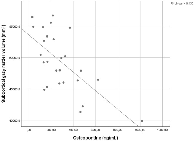
Osteopontin (OPN) is a proinflammatory marker produced by systemic immune and central nervous system (CNS) resident cells. We examined, if the level of OPN in the cerebrospinal fluid (CSF) and blood is associated with late-time regional brain volumes and white matter (WM) lesion load in MS. Concentrations of OPN in blood and CSF were related to MRI findings 10.1 ± 2.0 years later in 46 patients with MS. OPN concentration was measured by ELISA, while regional brain volumes and lesion load was assessed by magnetic resonance imaging (MRI) using 3D MPRAGE sequence and automated MR volumetry. OPN measured in the CSF was associated with several regional brain volumes and WM lesion load measured 10.1 ± 2.0 years later. CSF OPN concentration correlated with long-term enlargement of lateral- and inferior lateral ventricles and the elevation of gross CSF volume, in conjunction with the reduction of several cortical/subcortical gray matter and WM volumes. Serum OPN showed no long-term association with regional brain volumes. OPN measured from the CSF but not from the serum was associated with lower regional brain volumes measured a decade later, indicating the primary role of inflammation within the CNS in developing long-term brain related alterations.

摘要

骨桥蛋白 (OPN) 是一种由全身免疫细胞和中枢神经系统 (CNS) 固有细胞产生的促炎标志物。我们研究了脑脊液 (CSF) 和血液中的 OPN 水平是否与 MS 患者的晚期区域性脑容量和白质 (WM) 病变负荷有关。在 46 例 MS 患者中,OPN 在血液和 CSF 中的浓度与 10.1±2.0 年后的 MRI 发现相关。OPN 浓度通过 ELISA 测量,而区域性脑容量和病变负荷通过磁共振成像 (MRI) 使用 3D MPRAGE 序列和自动 MR 容积测量来评估。CSF 中测量的 OPN 与 10.1±2.0 年后测量的几个区域性脑容量和 WM 病变负荷相关。CSF OPN 浓度与侧脑室和下外侧脑室的长期扩大以及总 CSF 体积的升高相关,同时伴有几个皮质/皮质下灰质和 WM 体积的减少。血清 OPN 与区域性脑容量无长期关联。CSF 中测量的 OPN 而不是血清中测量的 OPN 与 10 年后测量的区域性脑容量较低有关,表明 CNS 内的炎症在发展长期与大脑相关的改变方面起主要作用。

https://cdn.ncbi.nlm.nih.gov/pmc/blobs/3f6a/8654976/2514e9794271/41598_2021_3173_Fig2_HTML.jpg
https://cdn.ncbi.nlm.nih.gov/pmc/blobs/3f6a/8654976/eb4a4d2d1415/41598_2021_3173_Fig1_HTML.jpg
https://cdn.ncbi.nlm.nih.gov/pmc/blobs/3f6a/8654976/2514e9794271/41598_2021_3173_Fig2_HTML.jpg
https://cdn.ncbi.nlm.nih.gov/pmc/blobs/3f6a/8654976/eb4a4d2d1415/41598_2021_3173_Fig1_HTML.jpg
https://cdn.ncbi.nlm.nih.gov/pmc/blobs/3f6a/8654976/2514e9794271/41598_2021_3173_Fig2_HTML.jpg

相似文献

1
Osteopontin levels are associated with late-time lower regional brain volumes in multiple sclerosis.骨桥蛋白水平与多发性硬化症晚期区域性脑容量降低有关。
Sci Rep. 2021 Dec 8;11(1):23604. doi: 10.1038/s41598-021-03173-3.
2
Microstructural and functional brain abnormalities in multiple sclerosis predicted by osteopontin and neurofilament light.骨桥蛋白和神经丝轻链预测多发性硬化症的脑微观结构和功能异常。
Mult Scler Relat Disord. 2021 Jun;51:102923. doi: 10.1016/j.msard.2021.102923. Epub 2021 Mar 24.
3
Cerebrospinal fluid neurofilament light levels mark grey matter volume in clinically isolated syndrome suggestive of multiple sclerosis.脑脊液神经丝轻链水平可标记临床孤立综合征中提示多发性硬化症的灰质体积。
Mult Scler. 2018 Jul;24(8):1039-1045. doi: 10.1177/1352458517711774. Epub 2017 May 24.
4
Osteopontin (OPN) as a CSF and blood biomarker for multiple sclerosis: A systematic review and meta-analysis.骨桥蛋白(OPN)作为多发性硬化症的脑脊液和血液生物标志物:系统评价与荟萃分析
PLoS One. 2018 Jan 18;13(1):e0190252. doi: 10.1371/journal.pone.0190252. eCollection 2018.
5
Increased levels of IL-23 and osteopontin in serum and cerebrospinal fluid of multiple sclerosis patients.多发性硬化症患者血清和脑脊液中白细胞介素-23 和骨桥蛋白水平升高。
J Neuroimmunol. 2012 Mar;244(1-2):94-6. doi: 10.1016/j.jneuroim.2011.12.004. Epub 2012 Feb 12.
6
Evaluation of circulating osteopontin levels in an unselected cohort of patients with multiple sclerosis: relevance for biomarker development.评估多发性硬化症未选择患者群中循环骨桥蛋白水平:对生物标志物开发的相关性。
Mult Scler. 2014 Apr;20(4):438-44. doi: 10.1177/1352458513503052. Epub 2013 Sep 4.
7
The predictive value of CSF multiple assay in multiple sclerosis: A single center experience.CSF 多项检测在多发性硬化症中的预测价值:单中心经验。
Mult Scler Relat Disord. 2019 Oct;35:176-181. doi: 10.1016/j.msard.2019.07.030. Epub 2019 Jul 28.
8
Osteopontin predicts late-time salience network-related functional connectivity in multiple sclerosis.骨桥蛋白预测多发性硬化症晚期突显网络相关功能连接。
PLoS One. 2024 Aug 29;19(8):e0309563. doi: 10.1371/journal.pone.0309563. eCollection 2024.
9
Association of Levels of CSF Osteopontin With Cortical Atrophy and Disability in Early Multiple Sclerosis.CSF 骨桥蛋白水平与早期多发性硬化症皮质萎缩和残疾的相关性研究。
Neurol Neuroimmunol Neuroinflamm. 2024 Sep;11(5):e200265. doi: 10.1212/NXI.0000000000200265. Epub 2024 Jun 25.
10
Characterization of gray-matter multiple sclerosis lesions using double inversion recovery, diffusion, contrast-enhanced, and volumetric MRI.采用双反转恢复、弥散、对比增强和容积 MRI 对灰质多发性硬化病变进行特征描述。
Mult Scler Relat Disord. 2019 Jun;31:74-81. doi: 10.1016/j.msard.2019.03.021. Epub 2019 Mar 29.

引用本文的文献

1
The Association of Axonal Damage Biomarkers and Osteopontin at Diagnosis Could Be Useful in Newly Diagnosed MS Patients.轴突损伤生物标志物与骨桥蛋白在诊断时的关联可能对新诊断的多发性硬化症患者有用。
Neurol Int. 2025 Jul 17;17(7):110. doi: 10.3390/neurolint17070110.
2
Fluid Biomarkers in Demyelinating Spectrum Disorders: Past, Present, and Prospects.脱髓鞘谱系障碍中的体液生物标志物:过去、现在与展望
Int J Mol Sci. 2025 May 7;26(9):4455. doi: 10.3390/ijms26094455.
3
Osteopontin predicts late-time salience network-related functional connectivity in multiple sclerosis.

本文引用的文献

1
Microstructural and functional brain abnormalities in multiple sclerosis predicted by osteopontin and neurofilament light.骨桥蛋白和神经丝轻链预测多发性硬化症的脑微观结构和功能异常。
Mult Scler Relat Disord. 2021 Jun;51:102923. doi: 10.1016/j.msard.2021.102923. Epub 2021 Mar 24.
2
Brain atrophy in multiple sclerosis: mechanisms, clinical relevance and treatment options.多发性硬化症中的脑萎缩:机制、临床相关性及治疗选择。
Auto Immun Highlights. 2019 Aug 10;10(1):7. doi: 10.1186/s13317-019-0117-5. eCollection 2019 Dec.
3
Advances in oral immunomodulating therapies in relapsing multiple sclerosis.
骨桥蛋白预测多发性硬化症晚期突显网络相关功能连接。
PLoS One. 2024 Aug 29;19(8):e0309563. doi: 10.1371/journal.pone.0309563. eCollection 2024.
4
Association of Levels of CSF Osteopontin With Cortical Atrophy and Disability in Early Multiple Sclerosis.CSF 骨桥蛋白水平与早期多发性硬化症皮质萎缩和残疾的相关性研究。
Neurol Neuroimmunol Neuroinflamm. 2024 Sep;11(5):e200265. doi: 10.1212/NXI.0000000000200265. Epub 2024 Jun 25.
5
New Enhancing MRI Lesions Associate with IL-17, Neutrophil Degranulation and Integrin Microparticles: Multi-Omics Combined with Frequent MRI in Multiple Sclerosis.新的增强型磁共振成像病变与白细胞介素-17、中性粒细胞脱颗粒和整合素微粒相关:多组学结合多发性硬化症的频繁磁共振成像
Biomedicines. 2023 Nov 28;11(12):3170. doi: 10.3390/biomedicines11123170.
6
Circulating microRNAs correlate with structural and functional MRI parameters in patients with multiple sclerosis.循环微小RNA与多发性硬化症患者的结构和功能磁共振成像参数相关。
Front Mol Neurosci. 2023 Oct 10;16:1173212. doi: 10.3389/fnmol.2023.1173212. eCollection 2023.
7
Proteomics and relationship with axonal pathology in multiple sclerosis: 5-year diffusion tensor imaging study.蛋白质组学与多发性硬化症轴突病理学的关系:5年扩散张量成像研究
Brain Commun. 2023 Jun 13;5(3):fcad183. doi: 10.1093/braincomms/fcad183. eCollection 2023.
8
Emerging imaging and liquid biomarkers in multiple sclerosis.多发性硬化症中的新兴影像和液体生物标志物。
Eur J Immunol. 2023 Aug;53(8):e2250228. doi: 10.1002/eji.202250228. Epub 2023 May 28.
9
Biomarkers in autoimmune diseases of the central nervous system.中枢神经系统自身免疫性疾病的生物标志物。
Front Immunol. 2023 Apr 5;14:1111719. doi: 10.3389/fimmu.2023.1111719. eCollection 2023.
10
Emerging Biomarkers of Multiple Sclerosis in the Blood and the CSF: A Focus on Neurofilaments and Therapeutic Considerations.血液和脑脊液中多发性硬化症的新兴生物标志物:聚焦神经丝蛋白及治疗考量
Int J Mol Sci. 2022 Mar 21;23(6):3383. doi: 10.3390/ijms23063383.
口服免疫调节疗法在复发型多发性硬化中的进展。
Lancet Neurol. 2020 Apr;19(4):336-347. doi: 10.1016/S1474-4422(19)30391-6. Epub 2020 Feb 11.
4
Molecular signature of different lesion types in the brain white matter of patients with progressive multiple sclerosis.不同病变类型在进展性多发性硬化症患者脑白质中的分子特征。
Acta Neuropathol Commun. 2019 Dec 11;7(1):205. doi: 10.1186/s40478-019-0855-7.
5
Pathogenic Mechanisms Associated With Different Clinical Courses of Multiple Sclerosis.与多发性硬化症不同临床病程相关的发病机制。
Front Immunol. 2019 Jan 10;9:3116. doi: 10.3389/fimmu.2018.03116. eCollection 2018.
6
Osteopontin (OPN) as a CSF and blood biomarker for multiple sclerosis: A systematic review and meta-analysis.骨桥蛋白(OPN)作为多发性硬化症的脑脊液和血液生物标志物:系统评价与荟萃分析
PLoS One. 2018 Jan 18;13(1):e0190252. doi: 10.1371/journal.pone.0190252. eCollection 2018.
7
Diagnosis of multiple sclerosis: 2017 revisions of the McDonald criteria.多发性硬化症的诊断:2017 年麦当劳标准修订版。
Lancet Neurol. 2018 Feb;17(2):162-173. doi: 10.1016/S1474-4422(17)30470-2. Epub 2017 Dec 21.
8
Osteopontin Augments M2 Microglia Response and Separates M1- and M2-Polarized Microglial Activation in Permanent Focal Cerebral Ischemia.骨桥蛋白增强 M2 小胶质细胞反应,并在永久性局灶性脑缺血中分离 M1 和 M2 极化的小胶质细胞激活。
Mediators Inflamm. 2017;2017:7189421. doi: 10.1155/2017/7189421. Epub 2017 Sep 20.
9
Correlation between white matter damage and gray matter lesions in multiple sclerosis patients.多发性硬化症患者白质损伤与灰质病变之间的相关性。
Neural Regen Res. 2017 May;12(5):787-794. doi: 10.4103/1673-5374.206650.
10
Cerebrospinal fluid neurofilament light levels mark grey matter volume in clinically isolated syndrome suggestive of multiple sclerosis.脑脊液神经丝轻链水平可标记临床孤立综合征中提示多发性硬化症的灰质体积。
Mult Scler. 2018 Jul;24(8):1039-1045. doi: 10.1177/1352458517711774. Epub 2017 May 24.