Wong Ying Mei, Jagmohan Pooja, Goh Yong Geng, Putti Thomas Choudary, Ow Samuel Guan Wei, Thian Yee Liang, Pillay Premilla
Department of Diagnostic Imaging, National University Hospital, Singapore, 1E Kent Ridge Road, NUHS Tower Block Level 12, Singapore, 119228, Singapore.
Department of Pathology, National University of Singapore, National University Hospital, Kent Ridge Road, Singapore, 119074, Singapore.
Insights Imaging. 2021 Dec 11;12(1):181. doi: 10.1186/s13244-021-01120-4.
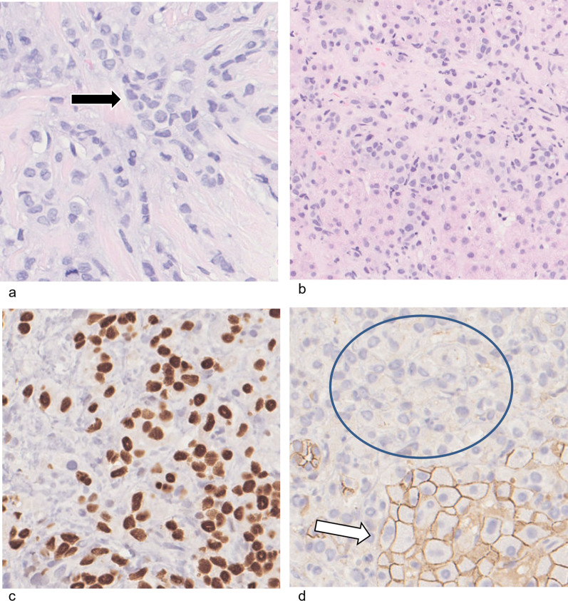
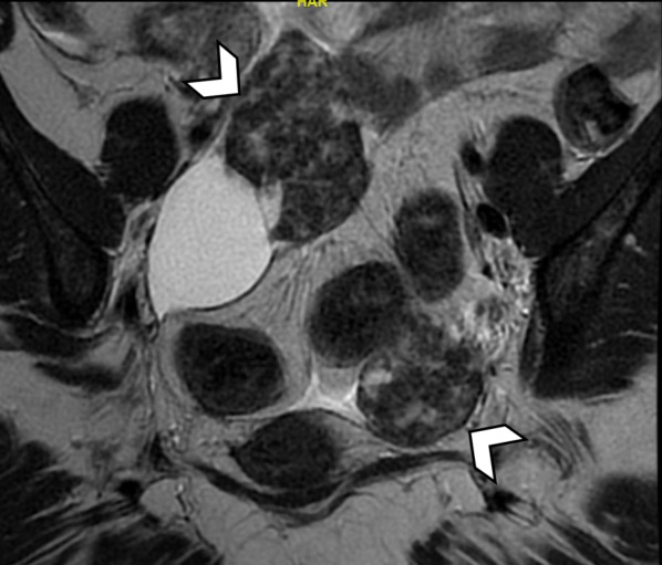
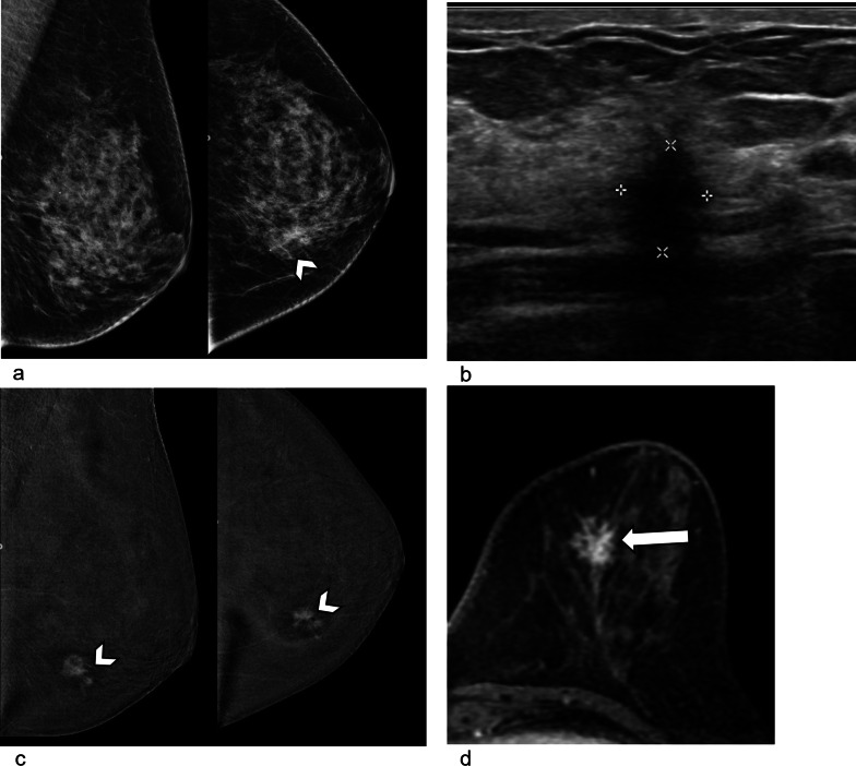
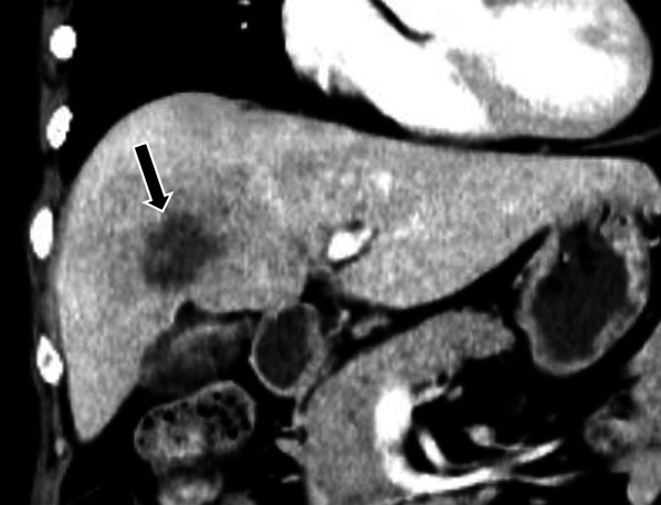
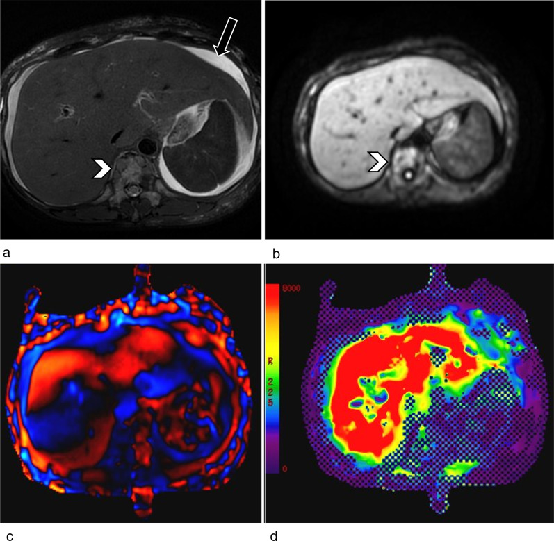
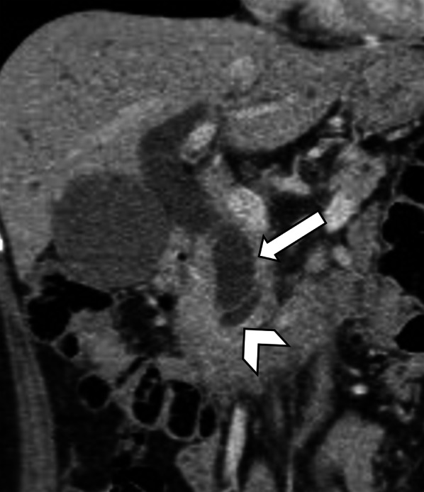
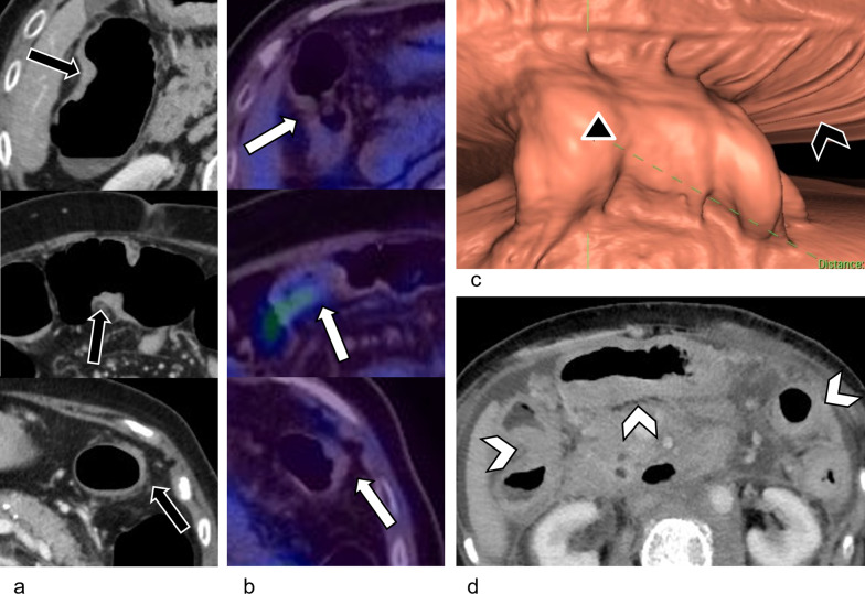
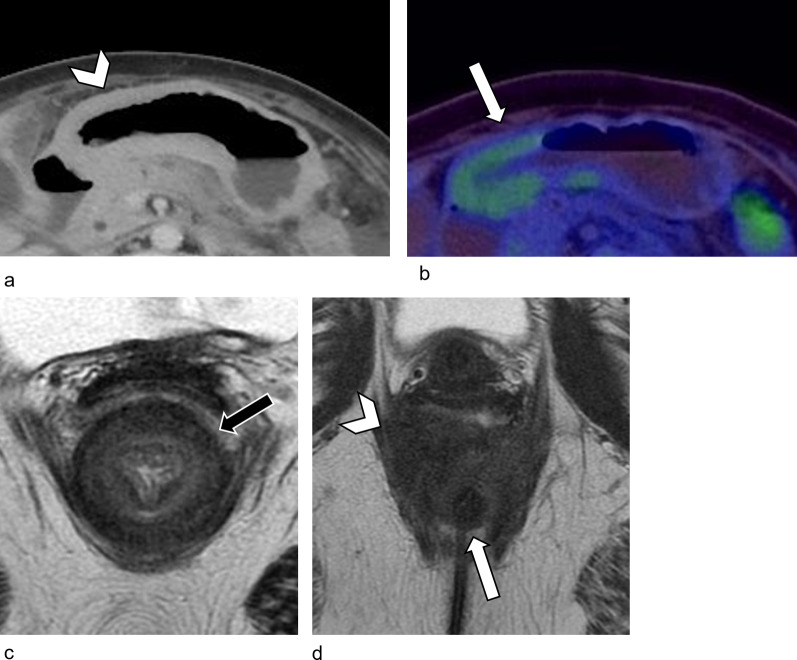
Invasive lobular carcinoma (ILC) has a greater tendency to metastasize to the peritoneum, retroperitoneum, and gastrointestinal (GI) tract as compared to invasive carcinoma of no special type (NST). Like primary ILC in the breast, ILC metastases are frequently infiltrative and hypometabolic, rather than mass forming and hypermetabolic in nature. This renders them difficult to detect on conventional and metabolic imaging studies. As a result, intra-abdominal ILC metastases are often detected late, with patients presenting with clinical complications such as liver failure, hydronephrosis, or bowel obstruction. In patients with known history of ILC, certain imaging features are very suggestive of infiltrative metastatic ILC. These include retroperitoneal or peritoneal nodularity and linitis plastica appearance of the bowel. Recognition of linitis plastica on imaging should prompt deep or repeat biopsies. In this pictorial review, the authors aim to familiarize readers with imaging features and pitfalls for evaluation of intra-abdominal metastatic ILC. Awareness of these will allow the radiologist to assess these patients with a high index of suspicion and aid detection of metastatic disease. Also, this can direct histopathology and immunohistochemical staining to obtain the correct diagnosis in suspected metastatic disease.
与非特殊类型浸润性癌(NST)相比,小叶原位癌(ILC)更容易转移至腹膜、腹膜后和胃肠道。与乳腺原发性ILC一样,ILC转移灶通常具有浸润性且代谢减低,而非形成肿块且代谢活跃。这使得它们在传统和代谢成像研究中难以检测到。因此,腹腔内ILC转移灶常常在晚期才被发现,患者表现出诸如肝衰竭、肾积水或肠梗阻等临床并发症。在有已知ILC病史的患者中,某些影像学特征非常提示浸润性转移性ILC。这些特征包括腹膜后或腹膜结节以及肠道的皮革胃表现。在影像学上识别皮革胃应促使进行深部或重复活检。在本图片综述中,作者旨在使读者熟悉评估腹腔内转移性ILC的影像学特征和陷阱。了解这些将使放射科医生以高度怀疑的态度评估这些患者,并有助于检测转移性疾病。此外,这可以指导组织病理学和免疫组化染色以在疑似转移性疾病中获得正确诊断。