• 文献检索
  • 文档翻译
  • 深度研究
  • 学术资讯
  • Suppr Zotero 插件Zotero 插件
  • 邀请有礼
  • 套餐&价格
  • 历史记录
应用&插件
Suppr Zotero 插件Zotero 插件浏览器插件Mac 客户端Windows 客户端微信小程序
定价
高级版会员购买积分包购买API积分包
服务
文献检索文档翻译深度研究API 文档MCP 服务
关于我们
关于 Suppr公司介绍联系我们用户协议隐私条款
关注我们

Suppr 超能文献

核心技术专利:CN118964589B侵权必究
粤ICP备2023148730 号-1Suppr @ 2026

文献检索

告别复杂PubMed语法,用中文像聊天一样搜索,搜遍4000万医学文献。AI智能推荐,让科研检索更轻松。

立即免费搜索

文件翻译

保留排版,准确专业,支持PDF/Word/PPT等文件格式,支持 12+语言互译。

免费翻译文档

深度研究

AI帮你快速写综述,25分钟生成高质量综述,智能提取关键信息,辅助科研写作。

立即免费体验

胎儿 18-24 孕周的心脏电轴:一项队列研究。

The electrical heart axis of the fetus between 18 and 24 weeks of gestation: A cohort study.

机构信息

Department of Obstetrics and Gynecology, Máxima Medical Center, Veldhoven, The Netherlands.

Eindhoven MedTech Innovation Center (e/MTIC), Eindhoven, The Netherlands.

出版信息

PLoS One. 2021 Dec 16;16(12):e0256115. doi: 10.1371/journal.pone.0256115. eCollection 2021.

DOI:10.1371/journal.pone.0256115
PMID:34914710
原文链接:https://pmc.ncbi.nlm.nih.gov/articles/PMC8675734/
Abstract

INTRODUCTION

A fetal anomaly scan in mid-pregnancy is performed, to check for the presence of congenital anomalies, including congenital heart disease (CHD). Unfortunately, 40% of CHD is still missed. The combined use of ultrasound and electrocardiography might boost detection rates. The electrical heart axis is one of the characteristics which can be deduced from an electrocardiogram (ECG). The aim of this study was to determine reference values for the electrical heart axis in healthy fetuses around 20 weeks of gestation.

MATERIAL AND METHODS

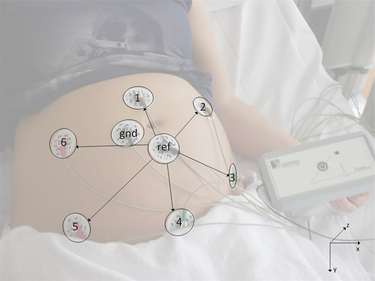
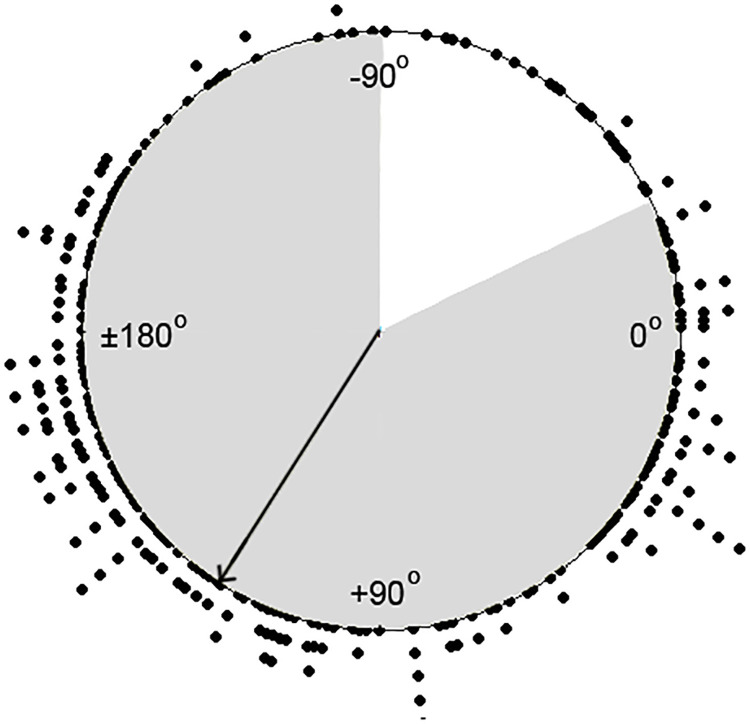
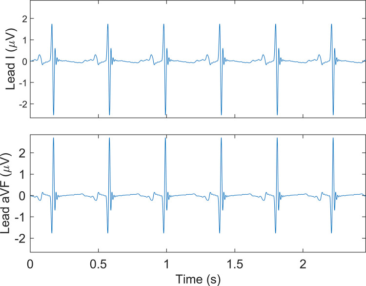
Non-invasive fetal electrocardiography was performed subsequent to the fetal anomaly scan in pregnant women carrying a healthy singleton fetus between 18 and 24 weeks of gestation. Eight adhesive electrodes were applied on the maternal abdomen including one ground and one reference electrode, yielding six channels of bipolar electrophysiological measurements. After removal of interferences, a fetal vectorcardiogram was calculated and then corrected for fetal orientation. The orientation of the electrical heart axis was determined from this normalized fetal vectorcardiogram. Descriptive statistics were used on normalized cartesian coordinates to determine the average electrical heart axis in the frontal plane. Furthermore, 90% prediction intervals (PI) for abnormality were calculated.

RESULTS

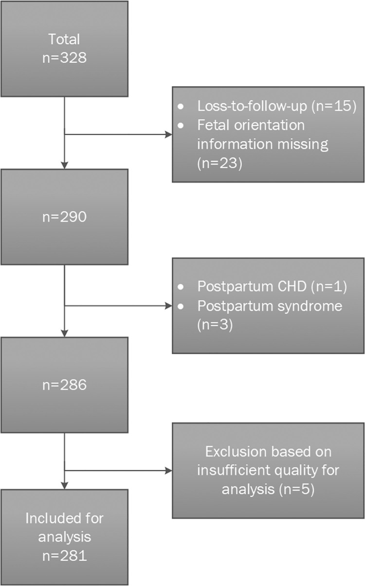
Of the 328 fetal ECGs performed, 281 were included in the analysis. The average electrical heart axis in the frontal plane was determined at 122.7° (90% PI: -25.6°; 270.9°).

DISCUSSION

The average electrical heart axis of healthy fetuses around mid-gestation is oriented to the right, which is, due to the unique fetal circulation, in line with muscle distribution in the fetal heart.

摘要

简介

在妊娠中期进行胎儿异常扫描,以检查是否存在先天性异常,包括先天性心脏病(CHD)。不幸的是,40%的 CHD 仍然会被漏诊。超声和心电图的联合使用可能会提高检测率。心脏电轴是可以从心电图(ECG)中推断出的特征之一。本研究旨在确定健康胎儿在妊娠 20 周左右的心脏电轴参考值。

材料和方法

在妊娠 18 至 24 周期间携带健康单胎的孕妇中,在进行胎儿异常扫描后,对胎儿进行非侵入性胎儿心电图检查。将 8 个粘性电极贴在孕妇腹部,包括一个接地电极和一个参考电极,产生 6 个双极电生理测量通道。去除干扰后,计算胎儿心向量图,然后根据胎儿方位进行校正。从这个正常化的胎儿心向量图中确定心脏电轴的方位。对正常化笛卡尔坐标进行描述性统计,以确定额面的平均心脏电轴。此外,还计算了异常的 90%预测区间(PI)。

结果

在进行的 328 次胎儿心电图中,有 281 次纳入分析。额面的平均心脏电轴确定为 122.7°(90%PI:-25.6°;270.9°)。

讨论

健康胎儿在妊娠中期的平均心脏电轴向右,这与独特的胎儿循环有关,与胎儿心脏中的肌肉分布一致。

https://cdn.ncbi.nlm.nih.gov/pmc/blobs/fc01/8675734/0bada49e7385/pone.0256115.g004.jpg
https://cdn.ncbi.nlm.nih.gov/pmc/blobs/fc01/8675734/804827d45dbe/pone.0256115.g001.jpg
https://cdn.ncbi.nlm.nih.gov/pmc/blobs/fc01/8675734/7630b48f236c/pone.0256115.g002.jpg
https://cdn.ncbi.nlm.nih.gov/pmc/blobs/fc01/8675734/d3643c8a9055/pone.0256115.g003.jpg
https://cdn.ncbi.nlm.nih.gov/pmc/blobs/fc01/8675734/0bada49e7385/pone.0256115.g004.jpg

相似文献

1
The electrical heart axis of the fetus between 18 and 24 weeks of gestation: A cohort study.胎儿 18-24 孕周的心脏电轴:一项队列研究。
PLoS One. 2021 Dec 16;16(12):e0256115. doi: 10.1371/journal.pone.0256115. eCollection 2021.
2
The standardized 12-lead fetal electrocardiogram of the healthy fetus in mid-pregnancy: A cross-sectional study.健康中期妊娠胎儿的标准化 12 导联胎儿心电图:一项横断面研究。
PLoS One. 2020 Apr 30;15(4):e0232606. doi: 10.1371/journal.pone.0232606. eCollection 2020.
3
The electrical heart axis in fetuses with congenital heart disease, measured with non-invasive fetal electrocardiography.先天性心脏病胎儿的无创性胎儿心电图测量的心脏电轴。
PLoS One. 2022 Oct 20;17(10):e0275802. doi: 10.1371/journal.pone.0275802. eCollection 2022.
4
Normal ranges for fetal electrocardiogram values for the healthy fetus of 18-24 weeks of gestation: a prospective cohort study.孕18 - 24周健康胎儿心电图值的正常范围:一项前瞻性队列研究。
BMC Pregnancy Childbirth. 2016 Aug 17;16:227. doi: 10.1186/s12884-016-1021-x.
5
Feasibility of non-invasive Foetal electrocardiography in a twin pregnancy.双胎妊娠中行胎儿无创心电图检查的可行性。
BMC Pregnancy Childbirth. 2020 Apr 15;20(1):215. doi: 10.1186/s12884-020-02918-8.
6
A systematic review of prenatal screening for congenital heart disease by fetal electrocardiography.胎儿心电图用于先天性心脏病产前筛查的系统评价
Int J Gynaecol Obstet. 2016 Nov;135(2):129-134. doi: 10.1016/j.ijgo.2016.05.010. Epub 2016 Aug 4.
7
Fetal cardiac axis in non-anomalous pregnancies: does fetal gender or maternal body mass index (BMI) matter?正常妊娠中的胎儿心脏轴:胎儿性别或孕妇体重指数(BMI)有影响吗?
J Matern Fetal Neonatal Med. 2016;29(13):2125-30. doi: 10.3109/14767058.2015.1077219. Epub 2015 Sep 12.
8
Fetal cardiac axis and congenital heart defects in early gestation.孕早期胎儿心脏轴与先天性心脏缺陷
Obstet Gynecol. 2015 Feb;125(2):453-460. doi: 10.1097/AOG.0000000000000608.
9
The Influence of Vectorcardiogram Orientation on the T/QRS Ratio Obtained Via Non-Invasive Fetal ECG.向量心电图方向对通过无创胎儿心电图获得的T/QRS比值的影响。
Annu Int Conf IEEE Eng Med Biol Soc. 2019 Jul;2019:1883-1886. doi: 10.1109/EMBC.2019.8857284.
10
Review of electromagnetic source investigations of the fetal heart.
Med Eng Phys. 2003 Dec;25(10):801-10. doi: 10.1016/s1350-4533(03)00121-8.

引用本文的文献

1
Fetal electrocardiography and artificial intelligence for prenatal detection of congenital heart disease.胎儿心电图与人工智能在先天性心脏病产前检测中的应用。
Acta Obstet Gynecol Scand. 2023 Nov;102(11):1511-1520. doi: 10.1111/aogs.14623. Epub 2023 Aug 10.
2
Adapting to a new environment: postnatal maturation of the human cardiomyocyte.适应新环境:人类心肌细胞的产后成熟。
J Physiol. 2023 Jul;601(13):2593-2619. doi: 10.1113/JP283792. Epub 2023 Apr 9.
3
The electrical heart axis in fetuses with congenital heart disease, measured with non-invasive fetal electrocardiography.

本文引用的文献

1
The standardized 12-lead fetal electrocardiogram of the healthy fetus in mid-pregnancy: A cross-sectional study.健康中期妊娠胎儿的标准化 12 导联胎儿心电图:一项横断面研究。
PLoS One. 2020 Apr 30;15(4):e0232606. doi: 10.1371/journal.pone.0232606. eCollection 2020.
2
The fetal electrocardiogram to detect the effects of betamethasone on fetal heart rate variability.检测倍他米松对胎儿心率变异性影响的胎儿心电图。
Early Hum Dev. 2019 Mar;130:57-64. doi: 10.1016/j.earlhumdev.2019.01.011. Epub 2019 Jan 22.
3
Antenatal Detection of Treatable Critical Congenital Heart Disease Is Associated with Lower Morbidity and Mortality.
先天性心脏病胎儿的无创性胎儿心电图测量的心脏电轴。
PLoS One. 2022 Oct 20;17(10):e0275802. doi: 10.1371/journal.pone.0275802. eCollection 2022.
产前检测可治疗的严重先天性心脏病与较低的发病率和死亡率相关。
J Pediatr. 2019 Jan;204:66-70. doi: 10.1016/j.jpeds.2018.08.056. Epub 2018 Oct 3.
4
Echocardiographic Evaluation of Ventricular Function-For the Neonatologist and Pediatric Intensivist.心室功能的超声心动图评估——面向新生儿科医生和儿科重症监护医生
Front Pediatr. 2018 Apr 4;6:79. doi: 10.3389/fped.2018.00079. eCollection 2018.
5
The impact of maternal obesity on completion of fetal anomaly screening.母亲肥胖对胎儿畸形筛查完成情况的影响。
J Perinat Med. 2017 Dec 20;45(9):1061-1067. doi: 10.1515/jpm-2016-0048.
6
Orientation of the electrical heart axis in mid-term pregnancy.妊娠中期心脏电轴的方向。
Eur J Obstet Gynecol Reprod Biol. 2016 Dec;207:243-246. doi: 10.1016/j.ejogrb.2016.10.030. Epub 2016 Oct 29.
7
Effect of tocolytic drugs on fetal heart rate variability: a systematic review.宫缩抑制药物对胎儿心率变异性的影响:一项系统评价
J Matern Fetal Neonatal Med. 2017 Oct;30(20):2387-2394. doi: 10.1080/14767058.2016.1249844. Epub 2016 Nov 8.
8
Normal ranges for fetal electrocardiogram values for the healthy fetus of 18-24 weeks of gestation: a prospective cohort study.孕18 - 24周健康胎儿心电图值的正常范围:一项前瞻性队列研究。
BMC Pregnancy Childbirth. 2016 Aug 17;16:227. doi: 10.1186/s12884-016-1021-x.
9
Prenatal detection of congenital heart disease--results of a national screening programme.先天性心脏病的产前检测——国家筛查计划的结果。
BJOG. 2016 Feb;123(3):400-7. doi: 10.1111/1471-0528.13274. Epub 2015 Jan 27.
10
The influence of maternal body mass index on fetal anomaly screening.孕妇体重指数对胎儿异常筛查的影响。
Eur J Obstet Gynecol Reprod Biol. 2014 Nov;182:181-4. doi: 10.1016/j.ejogrb.2014.09.031. Epub 2014 Sep 27.