Department of Radiological Sciences, University of California Irvine, 101 The City Dr S Building 55, Suite 201, Orange, CA, 92868, USA.
Department of Pathology and Laboratory Medicine, Medical Sciences I, D-440, University of California Irvine School of Medicine, Irvine, CA, 92697-4800, USA.
J Med Case Rep. 2021 Dec 17;15(1):597. doi: 10.1186/s13256-021-03183-9.
Pneumatosis cystoides intestinalis is a rare and usually benign condition in which multiple thin-walled cysts develop in the submucosa or subserosa of the gastrointestinal tract. While usually asymptomatic, severe cases can result in pneumoperitoneum, which can be managed surgically or medically depending on circumstances. We present a case of a patient with pneumatosis cystoides intestinalis, which presented as pneumoperitoneum following trauma. To our knowledge, there are no other published cases in which a trauma patient with pneumoperitoneum was found to have radiologic evidence of pneumatosis cystoides intestinalis.
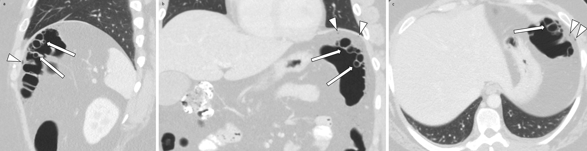
We present the case of a 37-year-old Hispanic male admitted to the hospital after being involved in a motorcycle accident. Computed tomography imaging of the abdomen and pelvis with oral and intravenous contrast demonstrated trace pneumoperitoneum, possibly originating from the splenic flexure of the colon without evidence of extravasation of oral contrast. Laparoscopy with conversion to exploratory laparotomy revealed bowel abnormalities at the distal transverse colon and splenic flexure, which were identified as pneumatosis cystoides intestinalis by pathology. There was no evidence of bowel perforation. A panel of abdominal radiologists attended the computed tomography interpretation to note that incidental atraumatic or traumatic rupture of the cysts could have caused the pneumoperitoneum. The patient had an uncomplicated postoperative course and was transferred to another facility per insurance request.
When presenting in the context of trauma, pneumatosis cystoides intestinalis can lead to difficult management decisions. To our knowledge, there are no existing evidence-based guidelines for the scenario of concurrent pneumatosis cystoides intestinalis, blunt abdominal trauma, and pneumoperitoneum in a patient with a benign abdominal exam. This patient's pneumoperitoneum was likely caused by rupture of preexisting cysts rather than frank bowel perforation. Patients who are asymptomatic, lack signs of clinically worrisome disease, and have a low pretest probability will likely not benefit from surgery and can be medically managed. Thorough discussion between surgeons and radiologists can be helpful when evaluating the clinical significance of a patient's pneumatosis cystoides intestinalis and aid in the decision to perform surgery.
肠气囊肿症是一种罕见且通常为良性的疾病,其特征为胃肠道黏膜下或浆膜下出现多个薄壁囊泡。虽然通常无症状,但严重的病例可导致气腹,根据具体情况,可通过手术或药物治疗进行管理。我们报告了一例肠气囊肿症患者,该患者在创伤后表现为气腹。据我们所知,目前尚无其他发表的病例报告表明创伤后气腹患者存在肠气囊肿症的放射学证据。
我们报告了一例 37 岁西班牙裔男性患者的病例,该患者在骑摩托车时发生事故后被收入院。腹部和骨盆的口服和静脉对比 CT 成像显示微量气腹,可能源自结肠脾曲,无口服造影剂外渗的证据。腹腔镜检查转为剖腹探查术显示横结肠远端和脾曲处的肠道异常,病理检查证实为肠气囊肿症。无肠穿孔的证据。一组腹部放射科医生参加了 CT 解读,指出偶然的无创伤性或创伤性的囊肿破裂可能导致气腹。患者术后恢复顺利,根据保险要求转往另一家机构。
当在创伤背景下出现时,肠气囊肿症可能导致困难的管理决策。据我们所知,对于同时存在肠气囊肿症、钝性腹部创伤和气腹且腹部检查为良性的患者,目前尚无基于现有证据的指南。该患者的气腹可能是由先前存在的囊肿破裂引起,而非真正的肠穿孔。无症状、缺乏有临床意义疾病表现且术前概率较低的患者可能不会从手术中获益,可进行药物治疗。在评估患者肠气囊肿症的临床意义并协助决定是否进行手术时,外科医生和放射科医生之间的充分讨论将有所帮助。