• 文献检索
  • 文档翻译
  • 深度研究
  • 学术资讯
  • Suppr Zotero 插件Zotero 插件
  • 邀请有礼
  • 套餐&价格
  • 历史记录
应用&插件
Suppr Zotero 插件Zotero 插件浏览器插件Mac 客户端Windows 客户端微信小程序
定价
高级版会员购买积分包购买API积分包
服务
文献检索文档翻译深度研究API 文档MCP 服务
关于我们
关于 Suppr公司介绍联系我们用户协议隐私条款
关注我们

Suppr 超能文献

核心技术专利:CN118964589B侵权必究
粤ICP备2023148730 号-1Suppr @ 2026

文献检索

告别复杂PubMed语法,用中文像聊天一样搜索,搜遍4000万医学文献。AI智能推荐,让科研检索更轻松。

立即免费搜索

文件翻译

保留排版,准确专业,支持PDF/Word/PPT等文件格式,支持 12+语言互译。

免费翻译文档

深度研究

AI帮你快速写综述,25分钟生成高质量综述,智能提取关键信息,辅助科研写作。

立即免费体验

使用三维MRI-CT图像融合图像对肘关节剥脱性骨软骨炎进行术前评估和手术模拟

Preoperative Evaluation and Surgical Simulation for Osteochondritis Dissecans of the Elbow Using Three-Dimensional MRI-CT Image Fusion Images.

作者信息

Kohyama Sho, Nishiura Yasumasa, Hara Yuki, Ogawa Takeshi, Ikumi Akira, Okano Eriko, Totoki Yasukazu, Yoshii Yuichi, Yamazaki Masashi

机构信息

Department of Orthopaedic Surgery, Kikkoman General Hospital, Noda 278-0005, Japan.

Tsuchiura Clinical Education and Training Center, Department of Orthopaedic Surgery, Tsukuba University Hospital, Tsuchiura 300-8585, Japan.

出版信息

Diagnostics (Basel). 2021 Dec 11;11(12):2337. doi: 10.3390/diagnostics11122337.

DOI:10.3390/diagnostics11122337
PMID:34943574
原文链接:https://pmc.ncbi.nlm.nih.gov/articles/PMC8700753/
Abstract

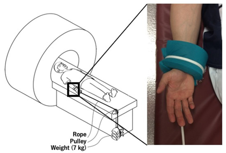
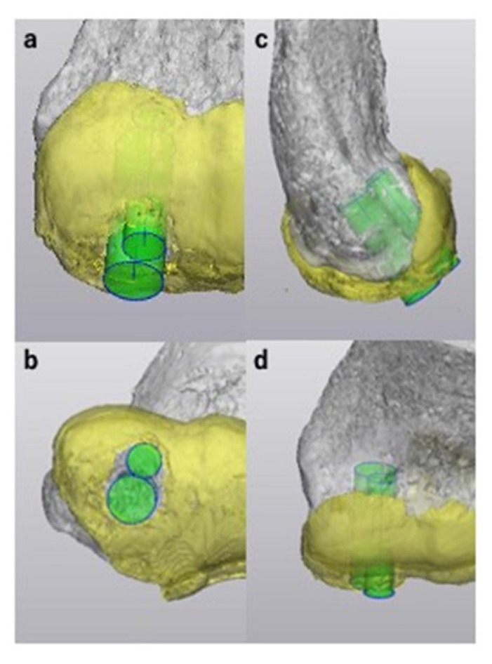
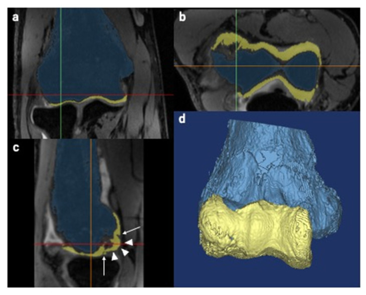
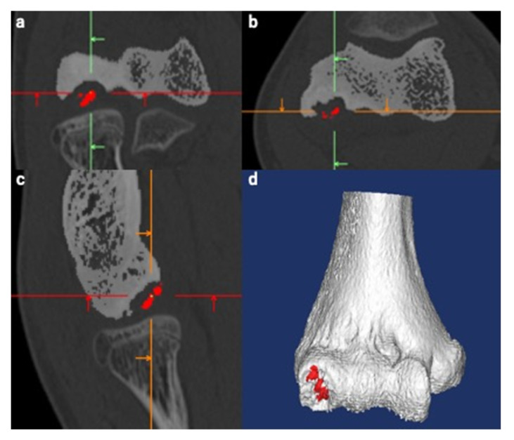
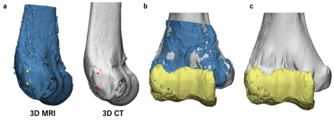
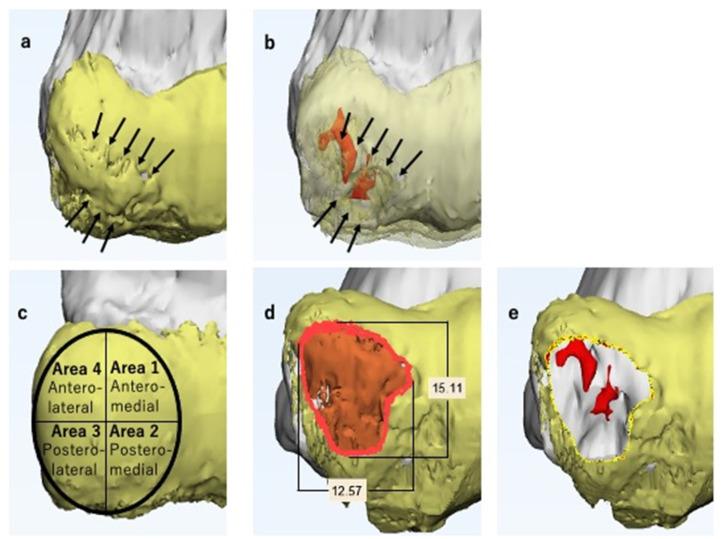
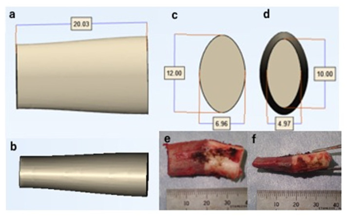
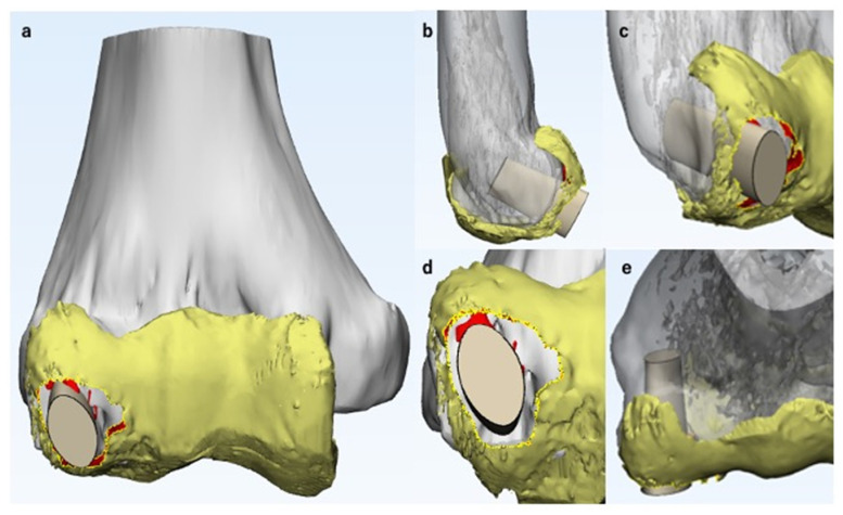
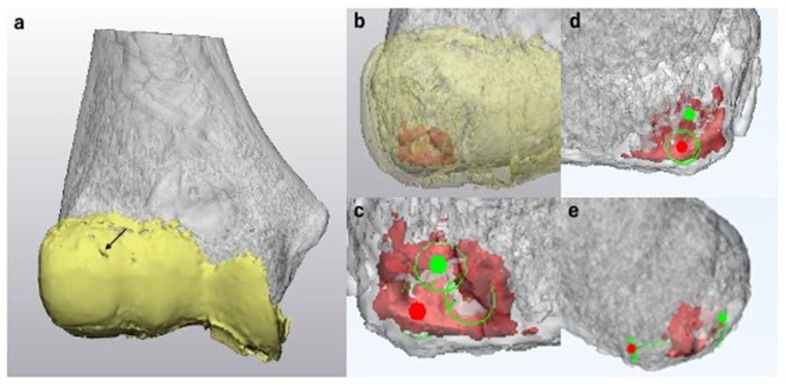
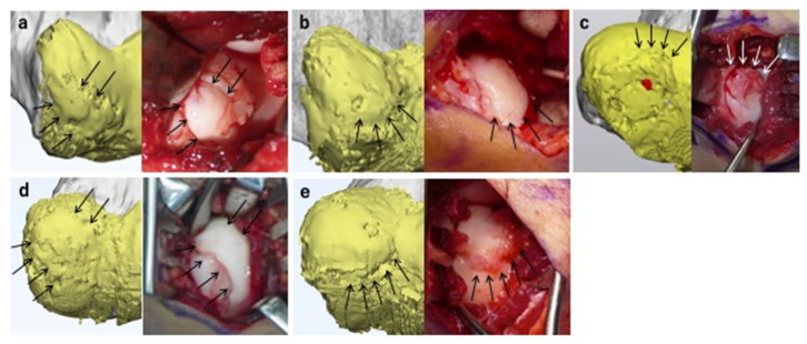
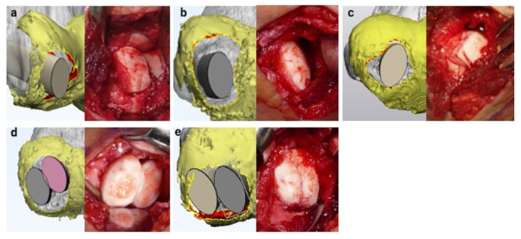
We used our novel three-dimensional magnetic resonance imaging-computed tomography fusion images (3D MRI-CT fusion images; MCFIs) for detailed preoperative lesion evaluation and surgical simulation in osteochondritis dissecans (OCD) of the elbow. Herein, we introduce our procedure and report the findings of the assessment of its utility. We enrolled 16 men (mean age: 14.0 years) and performed preoperative MRI using 7 kg axial traction with a 3-Tesla imager and CT. Three-dimensional-MRI models of the humerus and articular cartilage and a 3D-CT model of the humerus were constructed. We created MCFIs using both models. We validated the findings obtained from the MCFIs and intraoperative findings using the following items: articular cartilage fissures and defects, articular surface deformities, vertical and horizontal lesion diameters, the International Cartilage Repair Society (ICRS) classification, and surgical procedures. The MCFIs accurately reproduced the lesions and correctly matched the ICRS classification in 93.5% of cases. Surgery was performed as simulated in all cases. Preoperatively measured lesion diameters exhibited no significant differences compared to the intraoperative measurements. MCFIs were useful in the evaluation of OCD lesions and detailed preoperative surgical simulation through accurate reproduction of 3D structural details of the lesions.

摘要

我们使用新型三维磁共振成像 - 计算机断层扫描融合图像(3D MRI - CT融合图像;MCFIs)对肘关节剥脱性骨软骨炎(OCD)进行详细的术前病变评估和手术模拟。在此,我们介绍我们的操作流程并报告对其效用评估的结果。我们纳入了16名男性(平均年龄:14.0岁),使用3特斯拉成像仪和CT在7千克轴向牵引下进行术前MRI检查。构建了肱骨和关节软骨的三维MRI模型以及肱骨的三维CT模型。我们使用这两个模型创建了MCFIs。我们使用以下项目验证了从MCFIs获得的结果和术中发现:关节软骨裂隙和缺损、关节面畸形、病变的垂直和水平直径、国际软骨修复协会(ICRS)分类以及手术操作。MCFIs在93.5%的病例中准确再现了病变并正确匹配了ICRS分类。所有病例均按模拟进行手术。术前测量的病变直径与术中测量值相比无显著差异。MCFIs通过准确再现病变的三维结构细节,在OCD病变评估和详细的术前手术模拟中很有用。

https://cdn.ncbi.nlm.nih.gov/pmc/blobs/e5cc/8700753/2892e39606cb/diagnostics-11-02337-g011.jpg
https://cdn.ncbi.nlm.nih.gov/pmc/blobs/e5cc/8700753/ffaf9b858a7d/diagnostics-11-02337-g001.jpg
https://cdn.ncbi.nlm.nih.gov/pmc/blobs/e5cc/8700753/4d821cd3d819/diagnostics-11-02337-g002.jpg
https://cdn.ncbi.nlm.nih.gov/pmc/blobs/e5cc/8700753/a8ba7ebc6a6f/diagnostics-11-02337-g003.jpg
https://cdn.ncbi.nlm.nih.gov/pmc/blobs/e5cc/8700753/2e29fc5062a0/diagnostics-11-02337-g004.jpg
https://cdn.ncbi.nlm.nih.gov/pmc/blobs/e5cc/8700753/2b3512570289/diagnostics-11-02337-g005.jpg
https://cdn.ncbi.nlm.nih.gov/pmc/blobs/e5cc/8700753/dd904cc4702a/diagnostics-11-02337-g006.jpg
https://cdn.ncbi.nlm.nih.gov/pmc/blobs/e5cc/8700753/912a426a70fb/diagnostics-11-02337-g007.jpg
https://cdn.ncbi.nlm.nih.gov/pmc/blobs/e5cc/8700753/83e1965b082c/diagnostics-11-02337-g008.jpg
https://cdn.ncbi.nlm.nih.gov/pmc/blobs/e5cc/8700753/05d361958780/diagnostics-11-02337-g009.jpg
https://cdn.ncbi.nlm.nih.gov/pmc/blobs/e5cc/8700753/8e87500e7903/diagnostics-11-02337-g010.jpg
https://cdn.ncbi.nlm.nih.gov/pmc/blobs/e5cc/8700753/2892e39606cb/diagnostics-11-02337-g011.jpg

相似文献

1
Preoperative Evaluation and Surgical Simulation for Osteochondritis Dissecans of the Elbow Using Three-Dimensional MRI-CT Image Fusion Images.使用三维MRI-CT图像融合图像对肘关节剥脱性骨软骨炎进行术前评估和手术模拟
Diagnostics (Basel). 2021 Dec 11;11(12):2337. doi: 10.3390/diagnostics11122337.
2
A novel three-dimensional MRI-CT image fusion technique for precise preoperative evaluation and treatment of capitellar osteochondritis dissecans.一种用于精确术前评估和治疗肱骨小头剥脱性骨软骨炎的新型三维MRI-CT图像融合技术。
Eur Radiol. 2021 Aug;31(8):5721-5733. doi: 10.1007/s00330-020-07680-1. Epub 2021 Jan 16.
3
A Magnetic Resonance Imaging-Based Staging System for Osteochondritis Dissecans of the Elbow: A Validation Study Against the International Cartilage Repair Society Classification.基于磁共振成像的肘关节剥脱性骨软骨炎分期系统:针对国际软骨修复协会分类的验证研究
Orthop J Sports Med. 2018 Sep 12;6(9):2325967118794620. doi: 10.1177/2325967118794620. eCollection 2018 Sep.
4
Magnetic Resonance Imaging in Osteochondritis Dissecans of the Humeral Capitellum: Preoperative Assessment of Lesion Size and Lateral Wall Integrity.肱骨滑车骨软骨病损的磁共振成像:术前评估病变大小和外侧壁完整性。
J Hand Surg Am. 2021 Jun;46(6):454-461. doi: 10.1016/j.jhsa.2021.01.024. Epub 2021 Mar 29.
5
Magnetic Resonance Imaging of Osteochondritis Dissecans: Validation Study for the ICRS Classification System.剥脱性骨软骨炎的磁共振成像:国际软骨修复学会(ICRS)分类系统的验证研究
Acad Radiol. 2016 Jun;23(6):724-9. doi: 10.1016/j.acra.2016.01.015. Epub 2016 Mar 11.
6
Magnetic Resonance Imaging Staging to Evaluate the Stability of Capitellar Osteochondritis Dissecans Lesions.磁共振成像分期评估肱骨小头剥脱性骨软骨炎病变的稳定性
Am J Sports Med. 2014 Aug;42(8):1972-7. doi: 10.1177/0363546514532604. Epub 2014 May 9.
7
Comparison of sonography and MRI in the evaluation of stability of capitellar osteochondritis dissecans.超声与磁共振成像在评估肱骨小头剥脱性骨软骨炎稳定性中的比较。
J Clin Ultrasound. 2018 May;46(4):247-252. doi: 10.1002/jcu.22563. Epub 2017 Dec 6.
8
Results of Bone Peg Grafting for Capitellar Osteochondritis Dissecans in Adolescent Baseball Players.青少年棒球运动员肱骨小头剥脱性骨软骨炎的骨栓移植结果
Am J Sports Med. 2016 Dec;44(12):3171-3178. doi: 10.1177/0363546516658038. Epub 2016 Aug 11.
9
Preoperative imaging criteria for unstable osteochondritis dissecans of the capitellum.术前影像学标准在肱骨小头骺软骨分离不稳定中的应用。
Clin Orthop Relat Res. 2013 Apr;471(4):1137-43. doi: 10.1007/s11999-012-2462-9.
10
Autologous Osteochondral Mosaicplasty for Centrally and Laterally Located, Advanced Capitellar Osteochondritis Dissecans in Teenage Athletes: Clinical Outcomes, Radiography, and Magnetic Resonance Imaging Findings.自体骨软骨镶嵌移植术治疗青少年运动员中央和外侧型晚期肱骨滑车骨软骨炎:临床结果、影像学和磁共振成像表现。
Am J Sports Med. 2018 Jul;46(8):1943-1951. doi: 10.1177/0363546518768279. Epub 2018 May 8.

引用本文的文献

1
Osteochondritis Dissecans of the Elbow in Overhead Athletes: A Comprehensive Narrative Review.上肢运动员肘部剥脱性骨软骨炎:一项全面的叙述性综述
Diagnostics (Basel). 2024 Apr 28;14(9):916. doi: 10.3390/diagnostics14090916.
2
Advances in Bone Joint Imaging-Metal Artifact Reduction.骨关节成像——减少金属伪影的进展
Diagnostics (Basel). 2022 Dec 7;12(12):3079. doi: 10.3390/diagnostics12123079.
3
Use of a Small Car-Mounted Magnetic Resonance Imaging System for On-Field Screening for Osteochondritis Dissecans of the Humeral Capitellum.

本文引用的文献

1
Radiographic evaluation of osteochondritis dissecans of the humeral capitellum: A systematic review.肱骨小头剥脱性骨软骨炎的影像学评估:一项系统综述
J Orthop. 2021 Sep 14;27:114-121. doi: 10.1016/j.jor.2021.09.005. eCollection 2021 Sep-Oct.
2
Elbow arthroscopy - Indications and technique.肘关节镜检查——适应证与技术
J Clin Orthop Trauma. 2021 May 21;19:147-153. doi: 10.1016/j.jcot.2021.05.022. eCollection 2021 Aug.
3
The Use of Elbow Arthroscopy for Management of the Pediatric Elbow: A Systematic Review of Indications and Outcomes.
使用小型车载磁共振成像系统对肱骨小头剥脱性骨软骨炎进行现场筛查。
Diagnostics (Basel). 2022 Oct 20;12(10):2551. doi: 10.3390/diagnostics12102551.
4
Quantitative Assessment of the Restoration of Original Anatomy after 3D Virtual Reduction of Long Bone Fractures.长骨骨折三维虚拟复位后原始解剖结构恢复的定量评估。
Diagnostics (Basel). 2022 Jun 2;12(6):1372. doi: 10.3390/diagnostics12061372.
肘关镜在小儿肘部诊治中的应用:一项关于适应证和疗效的系统评价。
Arthroscopy. 2021 Jun;37(6):1958-1970.e1. doi: 10.1016/j.arthro.2021.01.047. Epub 2021 Feb 2.
4
A novel three-dimensional MRI-CT image fusion technique for precise preoperative evaluation and treatment of capitellar osteochondritis dissecans.一种用于精确术前评估和治疗肱骨小头剥脱性骨软骨炎的新型三维MRI-CT图像融合技术。
Eur Radiol. 2021 Aug;31(8):5721-5733. doi: 10.1007/s00330-020-07680-1. Epub 2021 Jan 16.
5
Effect of elbow MRI with axial traction on articular cartilage visibility-a feasibility study.肘关节 MRI 轴向牵引对关节软骨可视性的影响:一项可行性研究。
Skeletal Radiol. 2020 Oct;49(10):1555-1566. doi: 10.1007/s00256-020-03455-3. Epub 2020 May 4.
6
Capitellar Osteochondritis Dissecans Lesions of the Elbow: A Systematic Review of Osteochondral Graft Reconstruction Options.肘骨软骨骨软骨炎病变:骨软骨移植物重建选择的系统评价。
Arthroscopy. 2020 Jun;36(6):1747-1764. doi: 10.1016/j.arthro.2020.01.037. Epub 2020 Feb 6.
7
Osteochondritis Dissecans of the Elbow in Children: MRI Findings of Instability.儿童肘部剥脱性骨软骨炎:不稳定性的 MRI 表现。
AJR Am J Roentgenol. 2019 Nov;213(5):1145-1151. doi: 10.2214/AJR.19.21855. Epub 2019 Aug 28.
8
Osteochondritis Dissecans of Smaller Joints: The Elbow.小关节剥脱性骨软骨炎:肘。
Cartilage. 2021 Oct;12(4):407-417. doi: 10.1177/1947603519847735. Epub 2019 May 21.
9
Demographics and Epidemiology of Osteochondritis Dissecans of the Elbow Among Children and Adolescents.儿童和青少年肘部剥脱性骨软骨炎的人口统计学和流行病学
Orthop J Sports Med. 2018 Dec 19;6(12):2325967118815846. doi: 10.1177/2325967118815846. eCollection 2018 Dec.
10
Outcomes of surgical treatment for osteochondritis dissecans of the elbow: evaluation by lesion location.肘关节剥脱性骨软骨炎的手术治疗结果:根据病变位置进行评估。
J Shoulder Elbow Surg. 2018 Dec;27(12):2262-2270. doi: 10.1016/j.jse.2018.08.011.