• 文献检索
  • 文档翻译
  • 深度研究
  • 学术资讯
  • Suppr Zotero 插件Zotero 插件
  • 邀请有礼
  • 套餐&价格
  • 历史记录
应用&插件
Suppr Zotero 插件Zotero 插件浏览器插件Mac 客户端Windows 客户端微信小程序
定价
高级版会员购买积分包购买API积分包
服务
文献检索文档翻译深度研究API 文档MCP 服务
关于我们
关于 Suppr公司介绍联系我们用户协议隐私条款
关注我们

Suppr 超能文献

核心技术专利:CN118964589B侵权必究
粤ICP备2023148730 号-1Suppr @ 2026

文献检索

告别复杂PubMed语法,用中文像聊天一样搜索,搜遍4000万医学文献。AI智能推荐,让科研检索更轻松。

立即免费搜索

文件翻译

保留排版,准确专业,支持PDF/Word/PPT等文件格式,支持 12+语言互译。

免费翻译文档

深度研究

AI帮你快速写综述,25分钟生成高质量综述,智能提取关键信息,辅助科研写作。

立即免费体验

一种用于在培养的猪肾上皮细胞单层表面成像微绒毛生物发生的方案。

A protocol for imaging microvilli biogenesis on the surface of cultured porcine kidney epithelial cell monolayers.

机构信息

Department of Cell and Developmental Biology, Vanderbilt University, Nashville, TN 37232, USA.

Department of Medicine, Division of Infectious Diseases, Vanderbilt University Medical Center, Nashville, TN 37232, USA.

出版信息

STAR Protoc. 2021 Dec 10;2(4):100998. doi: 10.1016/j.xpro.2021.100998. eCollection 2021 Dec 17.

DOI:10.1016/j.xpro.2021.100998
PMID:34950883
原文链接:https://pmc.ncbi.nlm.nih.gov/articles/PMC8672049/
Abstract

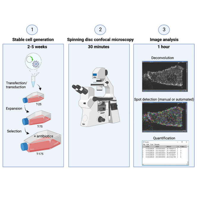
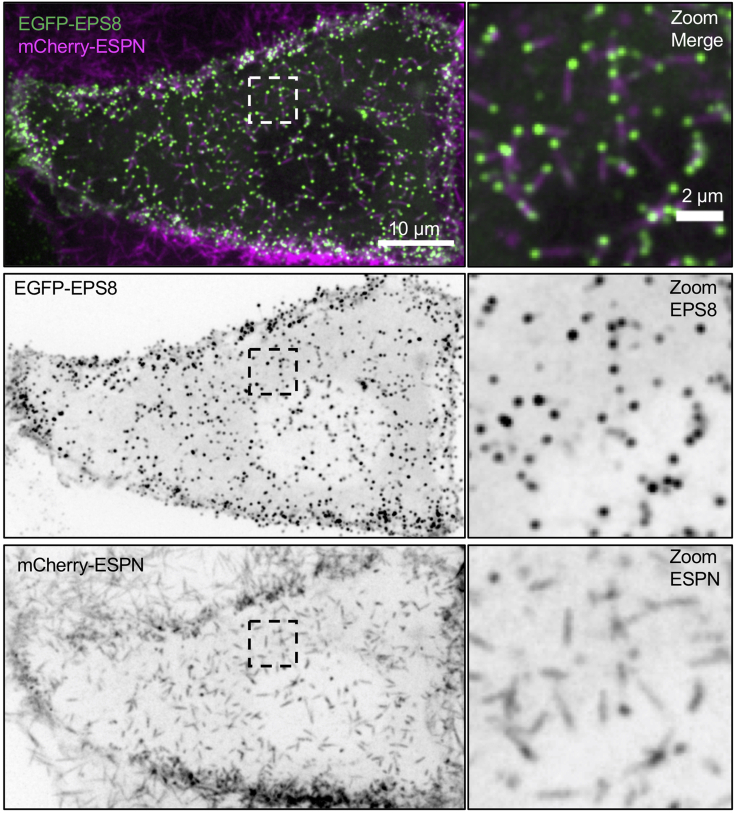
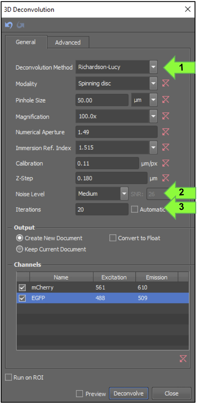
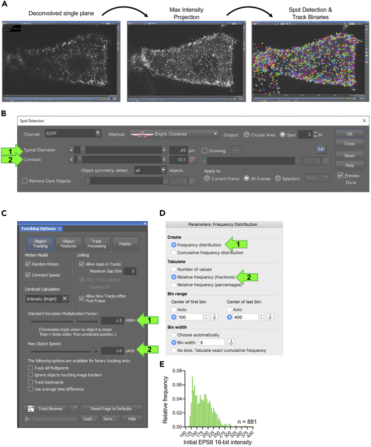
A key facet of epithelial differentiation is the assembly of actin-based protrusions known as microvilli, which amplify apical membrane surface area for various cell functions. To probe mechanisms of microvillus assembly, we developed a protocol using spinning disk confocal microscopy to directly visualize microvillus biogenesis on the surface of cultured porcine kidney epithelial cell monolayers engineered to express fluorescent proteins. This protocol offers access to the molecular details of individual protrusion growth events at high spatiotemporal resolution. For complete details on the use and execution of this protocol, please refer to Gaeta et al. (2021).

摘要

上皮细胞分化的一个关键方面是肌动蛋白为基础的突起的组装,这些突起被称为微绒毛,它们为各种细胞功能扩大了顶端膜表面积。为了探究微绒毛组装的机制,我们开发了一种使用旋转盘共聚焦显微镜的方案,该方案可直接在表达荧光蛋白的培养猪肾上皮细胞单层表面上可视化微绒毛的生物发生。该方案可在高时空分辨率下获得单个突起生长事件的分子细节。有关该方案的使用和执行的完整详细信息,请参阅 Gaeta 等人(2021 年)。

https://cdn.ncbi.nlm.nih.gov/pmc/blobs/5d7b/8672049/0eadcfe5ea1f/gr8.jpg
https://cdn.ncbi.nlm.nih.gov/pmc/blobs/5d7b/8672049/7ed8e61e8bc8/fx1.jpg
https://cdn.ncbi.nlm.nih.gov/pmc/blobs/5d7b/8672049/57c2ffeec534/gr1.jpg
https://cdn.ncbi.nlm.nih.gov/pmc/blobs/5d7b/8672049/cfcf957535fc/gr2.jpg
https://cdn.ncbi.nlm.nih.gov/pmc/blobs/5d7b/8672049/19181b4fab21/gr3.jpg
https://cdn.ncbi.nlm.nih.gov/pmc/blobs/5d7b/8672049/a16122c5f86d/gr5.jpg
https://cdn.ncbi.nlm.nih.gov/pmc/blobs/5d7b/8672049/80b2e477c30f/gr4.jpg
https://cdn.ncbi.nlm.nih.gov/pmc/blobs/5d7b/8672049/a24e90c18778/gr6.jpg
https://cdn.ncbi.nlm.nih.gov/pmc/blobs/5d7b/8672049/70db01d3e0e1/gr7.jpg
https://cdn.ncbi.nlm.nih.gov/pmc/blobs/5d7b/8672049/0eadcfe5ea1f/gr8.jpg

相似文献

1
A protocol for imaging microvilli biogenesis on the surface of cultured porcine kidney epithelial cell monolayers.一种用于在培养的猪肾上皮细胞单层表面成像微绒毛生物发生的方案。
STAR Protoc. 2021 Dec 10;2(4):100998. doi: 10.1016/j.xpro.2021.100998. eCollection 2021 Dec 17.
2
Direct visualization of epithelial microvilli biogenesis.上皮微绒毛生物发生的直接可视化。
Curr Biol. 2021 Jun 21;31(12):2561-2575.e6. doi: 10.1016/j.cub.2021.04.012. Epub 2021 May 4.
3
Selective localization of the polytopic membrane protein prominin in microvilli of epithelial cells - a combination of apical sorting and retention in plasma membrane protrusions.多拓扑膜蛋白prominin在上皮细胞微绒毛中的选择性定位——顶端分选与质膜突起中保留的结合。
J Cell Sci. 1999 Apr;112 ( Pt 7):1023-33. doi: 10.1242/jcs.112.7.1023.
4
Actin Dynamics Drive Microvillar Motility and Clustering during Brush Border Assembly.肌动蛋白动力学驱动微绒毛在刷状缘组装过程中的运动和聚集。
Dev Cell. 2019 Sep 9;50(5):545-556.e4. doi: 10.1016/j.devcel.2019.07.008. Epub 2019 Aug 1.
5
A protocol to quantify chromatin compaction with confocal and super-resolution microscopy in cultured cells.一种使用共聚焦和超分辨率显微镜在培养细胞中定量染色质紧缩的方案。
STAR Protoc. 2021 Sep 30;2(4):100865. doi: 10.1016/j.xpro.2021.100865. eCollection 2021 Dec 17.
6
Building the brush border, one microvillus at a time.一次一个微绒毛,构建刷状缘。
Curr Opin Cell Biol. 2023 Feb;80:102153. doi: 10.1016/j.ceb.2023.102153. Epub 2023 Feb 22.
7
Localization of capping protein in chicken epithelial cells by immunofluorescence and biochemical fractionation.通过免疫荧光和生化分级分离法对鸡上皮细胞中封端蛋白进行定位
J Cell Biol. 1992 Jul;118(2):335-46. doi: 10.1083/jcb.118.2.335.
8
Mitotic Spindle Positioning (MISP) is an actin bundler that selectively stabilizes the rootlets of epithelial microvilli.有丝分裂纺锤体定位(MISP)是一种肌动蛋白束形成蛋白,它选择性地稳定上皮微绒毛的根丝。
Cell Rep. 2022 Apr 19;39(3):110692. doi: 10.1016/j.celrep.2022.110692.
9
Correlative confocal and scanning electron microscopy of cultured cells without using dedicated equipment.培养细胞的无专用设备共聚焦和扫描电子显微镜相关观察。
STAR Protoc. 2021 Aug 9;2(3):100727. doi: 10.1016/j.xpro.2021.100727. eCollection 2021 Sep 17.
10
Protocol for the detection of mechanosensitive response of proteins in cultured cells under compressive stress.在压缩应力下培养细胞中机械敏感蛋白反应的检测方案。
STAR Protoc. 2024 Jun 21;5(2):103098. doi: 10.1016/j.xpro.2024.103098. Epub 2024 May 31.

引用本文的文献

1
Adhesion-based capture stabilizes nascent microvilli at epithelial cell junctions.基于黏附的捕获作用可稳定上皮细胞连接处的初生微绒毛。
Dev Cell. 2023 Oct 23;58(20):2048-2062.e7. doi: 10.1016/j.devcel.2023.09.001. Epub 2023 Oct 12.
2
BioID2 screening identifies KIAA1671 as an EPS8 proximal factor that marks sites of microvillus growth.BioID2 筛选鉴定出 KIAA1671 是 EPS8 近端因子,可作为微绒毛生长部位的标志物。
Mol Biol Cell. 2023 Apr 1;34(4):ar31. doi: 10.1091/mbc.E22-11-0498. Epub 2023 Feb 15.

本文引用的文献

1
Direct visualization of epithelial microvilli biogenesis.上皮微绒毛生物发生的直接可视化。
Curr Biol. 2021 Jun 21;31(12):2561-2575.e6. doi: 10.1016/j.cub.2021.04.012. Epub 2021 May 4.
2
Actin Dynamics Drive Microvillar Motility and Clustering during Brush Border Assembly.肌动蛋白动力学驱动微绒毛在刷状缘组装过程中的运动和聚集。
Dev Cell. 2019 Sep 9;50(5):545-556.e4. doi: 10.1016/j.devcel.2019.07.008. Epub 2019 Aug 1.
3
IRTKS (BAIAP2L1) Elongates Epithelial Microvilli Using EPS8-Dependent and Independent Mechanisms.IRTKS(BAIAP2L1)通过依赖和不依赖 EPS8 的机制来延长上皮微绒毛。
Curr Biol. 2018 Sep 24;28(18):2876-2888.e4. doi: 10.1016/j.cub.2018.07.022. Epub 2018 Sep 6.
4
Actin visualization at a glance.肌动蛋白可视化一览。
J Cell Sci. 2017 Feb 1;130(3):525-530. doi: 10.1242/jcs.189068. Epub 2017 Jan 12.
5
Culturing MDCK cells in three dimensions for analyzing intracellular dynamics.在三维环境中培养MDCK细胞以分析细胞内动力学。
Curr Protoc Cell Biol. 2009 Jun;Chapter 4:4.22.1-4.22.18. doi: 10.1002/0471143030.cb0422s43.
6
Complete polarization of single intestinal epithelial cells upon activation of LKB1 by STRAD.STRAD激活LKB1后单个肠上皮细胞的完全极化
Cell. 2004 Feb 6;116(3):457-66. doi: 10.1016/s0092-8674(04)00114-x.
7
MYO1A (brush border myosin I) dynamics in the brush border of LLC-PK1-CL4 cells.LLC-PK1-CL4细胞刷状缘中的MYO1A(刷状缘肌球蛋白I)动力学
Biophys J. 2002 Apr;82(4):1869-83. doi: 10.1016/S0006-3495(02)75537-9.
8
Characterization of the enterocyte-like brush border cytoskeleton of the C2BBe clones of the human intestinal cell line, Caco-2.
J Cell Sci. 1992 Jul;102 ( Pt 3):581-600. doi: 10.1242/jcs.102.3.581.