Department of Cell and Developmental Biology, Vanderbilt University School of Medicine, Nashville, TN 37232.
Mol Biol Cell. 2023 Apr 1;34(4):ar31. doi: 10.1091/mbc.E22-11-0498. Epub 2023 Feb 15.
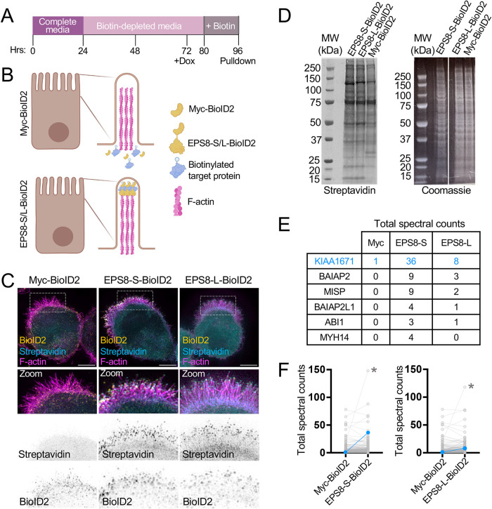
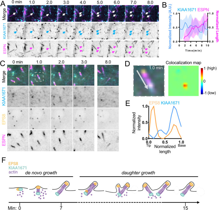
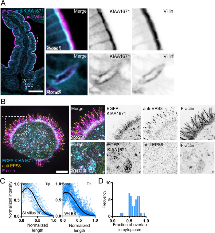
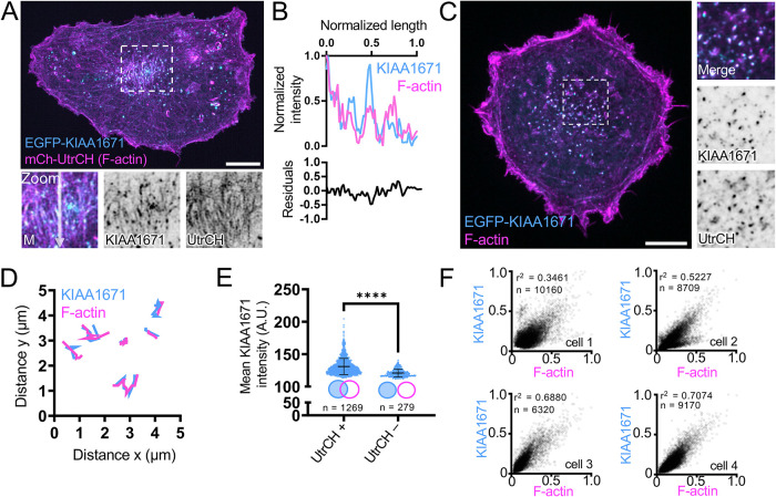
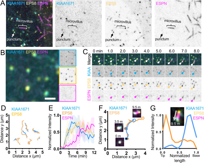
Microvilli are defining morphological features of the apical surfaces in diverse epithelial tissues. To develop our understanding of microvillus biogenesis, we used a biotin proximity-labeling approach to uncover new molecules enriched near EPS8, a well-studied marker of the microvillus distal tip compartment. Mass spectrometry of biotinylated hits identified KIAA1671, a large (∼200 kDa), disordered, and previously uncharacterized protein. Based on immunofluorescent staining and expression of fluorescent protein-tagged constructs, we found that KIAA1671 localizes to the base of the brush border in native intestinal tissue and polarized epithelial-cell culture models, as well as dynamic actin-rich structures in unpolarized, nonepithelial cell types. Live imaging also revealed that during the early stages of microvillar growth, KIAA1671 colocalizes with EPS8 in diffraction-limited puncta. However, once elongation of the core bundle begins, these two factors separate, with EPS8 tracking the distal end and KIAA1671 remaining behind at the base of the structure. These results suggest that KIAA1671 cooperates with EPS8 and potentially other assembly factors to initiate growth of microvilli on the apical surface. These findings offer new details on how transporting epithelial cells builds the brush border and may inform our understanding of how apical specializations are assembled in other epithelial contexts.
微绒毛是多种上皮组织中顶端表面的特有形态特征。为了深入了解微绒毛的生物发生过程,我们采用生物素邻近标记方法,揭示了新的靠近 EPS8 富集的分子,EPS8 是微绒毛远端尖端隔室的一个研究充分的标志物。生物素标记物的质谱分析鉴定出 KIAA1671,这是一种大型(∼200 kDa)、无序且以前未被表征的蛋白质。基于免疫荧光染色和荧光蛋白标记的构建体的表达,我们发现 KIAA1671 定位于天然肠组织和极化上皮细胞培养模型的刷状缘基底,以及非极化、非上皮细胞类型中的动态肌动蛋白丰富结构。活细胞成像还揭示了在微绒毛生长的早期阶段,KIAA1671 与 EPS8 在衍射限制的小点中共定位。然而,一旦核心束的伸长开始,这两个因素就会分离,EPS8 追踪远端,而 KIAA1671 留在结构的底部。这些结果表明 KIAA1671 与 EPS8 合作,并可能与其他组装因子一起,在上皮组织的顶端表面启动微绒毛的生长。这些发现提供了有关转运上皮细胞如何构建刷状缘的新细节,并可能有助于我们理解其他上皮组织中如何组装顶端特化结构。