State Key Laboratory of Oral Diseases, West China Hospital of Stomatology, Sichuan University, Chengdu, China.
Department of Orthodontics, West China Hospital of Stomatology, Sichuan University, Chengdu, China.
Cell Prolif. 2022 Jan;55(1):e13172. doi: 10.1111/cpr.13172. Epub 2021 Dec 24.
Aging and common diseases alter the stiffness of bone tissue, causing changes to the microenvironment of the mechanosensitive bone cells. Osteoclasts, the sole bone-resorbing cells, play a vital role in bone remodeling. This study was performed to elucidate the mechanism through which osteoclasts sense and react to substrate stiffness signals.
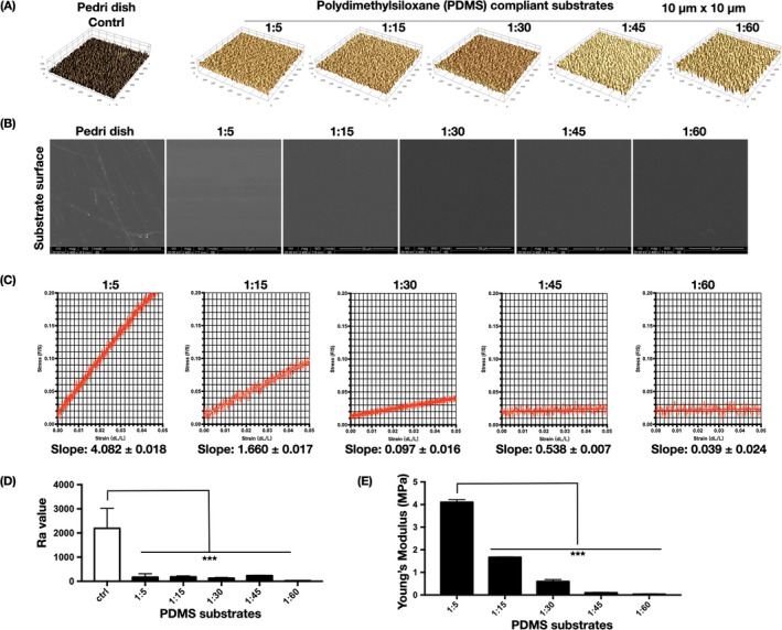
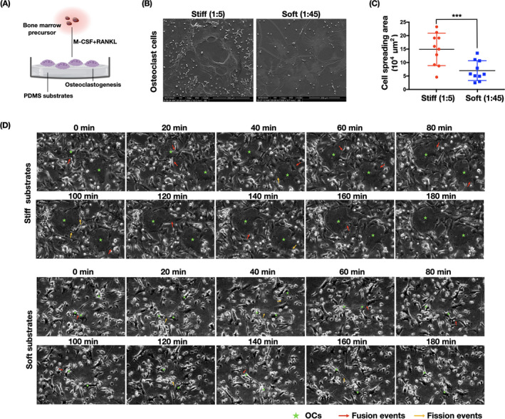
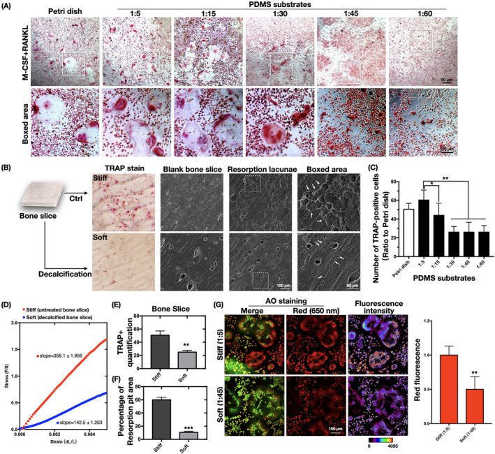
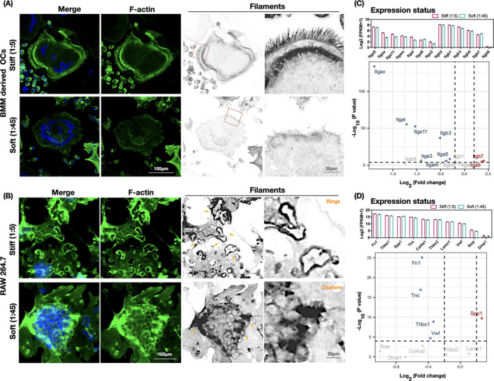
We fabricated polydimethylsiloxane (PDMS) substrates of different stiffness degrees for osteoclast formation progressed from osteoclast precursors including bone marrow-derived macrophages (BMMs) and RAW264.7 monocytes. Osteoclast differentiation in response to the stiffness signals was determined by examining the cell morphology, fusion/fission activities, transcriptional profile, and resorption function. Cytoskeletal changes and mechanosensitive adhesion molecules were also assessed.
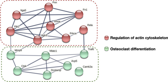
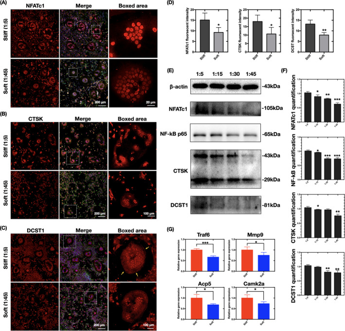
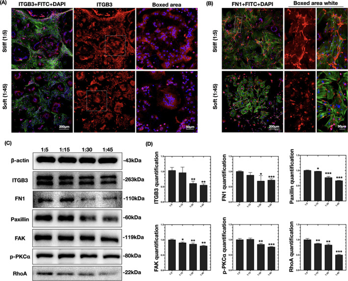
Stiffer PDMS substrates accelerated osteoclast differentiation, firstly observed by variations in their morphology and fusion/fission activities. Upregulation of canonical osteoclast markers (Nfatc1, Acp5, Ctsk, Camk2a, Mmp9, Rela, and Traf6) and the fusion master regulator DC-stamp were detected on stiffer substrates, with similar increases in their bone resorption functions. Additionally, the activation of cytoskeleton-associated adhesion molecules, including fibronectin and integrin αvβ3, followed by biochemical signaling cascades of paxillin, FAK, PKC, and RhoA, was detected on the stiffer substrates.
This is the first study to provide evidence proving that extracellular substrate stiffness is a strong determinant of osteoclast differentiation and functions. Higher stiffness upregulated the differentiation profile and activity of osteoclasts, revealing the mechanical regulation of osteoclast activity in bone homeostasis and diseases.
衰老和常见疾病会改变骨组织的硬度,导致机械敏感的破骨细胞的微环境发生变化。破骨细胞是唯一的骨吸收细胞,在骨重塑中起着至关重要的作用。本研究旨在阐明破骨细胞感知和反应基质硬度信号的机制。
我们制备了不同硬度的聚二甲基硅氧烷(PDMS)基质,用于从包括骨髓来源巨噬细胞(BMMs)和 RAW264.7 单核细胞在内的破骨细胞前体中进行破骨细胞形成。通过观察细胞形态、融合/裂变活性、转录谱和吸收功能来确定对刚度信号的破骨细胞分化。还评估了细胞骨架变化和机械敏感粘附分子。
更硬的 PDMS 基质加速了破骨细胞分化,首先通过其形态和融合/裂变活性的变化观察到。在更硬的基质上检测到经典破骨细胞标志物(Nfatc1、Acp5、Ctsk、Camk2a、Mmp9、Rela 和 Traf6)和融合主调节剂 DC-stamp 的上调,其骨吸收功能也相似增加。此外,在更硬的基质上检测到细胞骨架相关粘附分子(包括纤连蛋白和整合素 αvβ3)的激活,随后是细胞骨架相关粘附分子(包括纤连蛋白和整合素 αvβ3)的生化信号级联反应,包括桩蛋白、FAK、PKC 和 RhoA。
这是第一项提供证据证明细胞外基质硬度是破骨细胞分化和功能的重要决定因素的研究。更高的硬度上调了破骨细胞的分化特征和活性,揭示了骨稳态和疾病中破骨细胞活性的机械调节。