Multidisciplinary Group in Oncology (GERCOR), Paris, France.
Department of Surgical and Medical Oncology, University Hospital of Clermont-Ferrand, France.
Mol Oncol. 2022 Jun;16(11):2260-2273. doi: 10.1002/1878-0261.13173. Epub 2022 Feb 9.
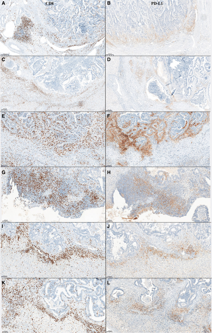
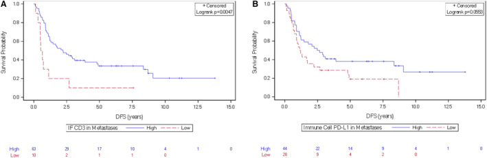
In the era of immune checkpoint inhibitors, understanding the metastatic microenvironment of proficient mismatch repair/microsatellite stable (pMMR/MSS) colorectal cancer (CRC) is of paramount importance to both prognostication and the development of more effective novel therapies. In this study, primary and paired metastasis tissue samples were collected from patients with resectable metastatic CRC treated with adjuvant FOLFOX or peri-operative chemotherapy in the MIROX phase III prospective study. In total, 74 cancer tissues were stained for CD3, CD8, Forkhead box protein 3 (FOXP3), programmed cell death protein-1 (PD-1, invasive front, stromal, intra-epithelial compartments), and programmed death-ligand 1 (PD-L1, tumor, immune cells). The immune profiling of primary CRC had a limited value to predict the immune context of paired metastases for all markers but CD3+. The expression of CD8 and PD-L1 was higher in metastases after neoadjuvant FOLFOX. In metastases, both CD3 T cells at the invasive front and PD-L1 expressions on immune cells were predictive of better disease-free survival. These results show that the effect of FOLFOX on modifying the immune microenvironment in resected CRC metastases and measurement of PD-L1 expression and tumor-infiltrating CD8 T cells in pMMR/MSS metastatic tissue samples could improve treatment strategies of metastatic CRC patients.
在免疫检查点抑制剂时代,了解熟练错配修复/微卫星稳定(pMMR/MSS)结直肠癌(CRC)的转移性微环境对于预后和开发更有效的新型治疗方法至关重要。在这项研究中,从接受辅助 FOLFOX 或围手术期化疗治疗的可切除转移性 CRC 患者中收集了原发和配对转移组织样本,这些患者来自 MIROX 三期前瞻性研究。总共对 74 个癌症组织进行了 CD3、CD8、叉头框蛋白 3(FOXP3)、程序性细胞死亡蛋白 1(PD-1,侵袭前沿、基质、上皮内隔室)和程序性死亡配体 1(PD-L1,肿瘤、免疫细胞)的染色。原发 CRC 的免疫分析对于预测所有标志物但 CD3+的配对转移的免疫背景的价值有限。新辅助 FOLFOX 后转移中 CD8 和 PD-L1 的表达更高。在转移中,侵袭前沿的 CD3 T 细胞和免疫细胞上的 PD-L1 表达均与无病生存时间更长相关。这些结果表明,FOLFOX 对切除的 CRC 转移中免疫微环境的修饰作用以及在 pMMR/MSS 转移性组织样本中测量 PD-L1 表达和肿瘤浸润性 CD8 T 细胞,可能改善转移性 CRC 患者的治疗策略。