Yeh Shin-Joe, Hsu Pang-Hung, Yeh Ti-Yen, Yang Wei-Kang, Chang Ko-Ping, Chiang Chien-Sung, Tang Sung-Chun, Tsai Li-Kai, Jeng Jiann-Shing, Hsieh Sung-Tsang
Graduate Institute of Anatomy and Cell Biology, National Taiwan University College of Medicine, Taipei, Taiwan.
Department of Neurology, National Taiwan University Hospital, Taipei, Taiwan.
Front Mol Neurosci. 2021 Dec 16;14:754762. doi: 10.3389/fnmol.2021.754762. eCollection 2021.
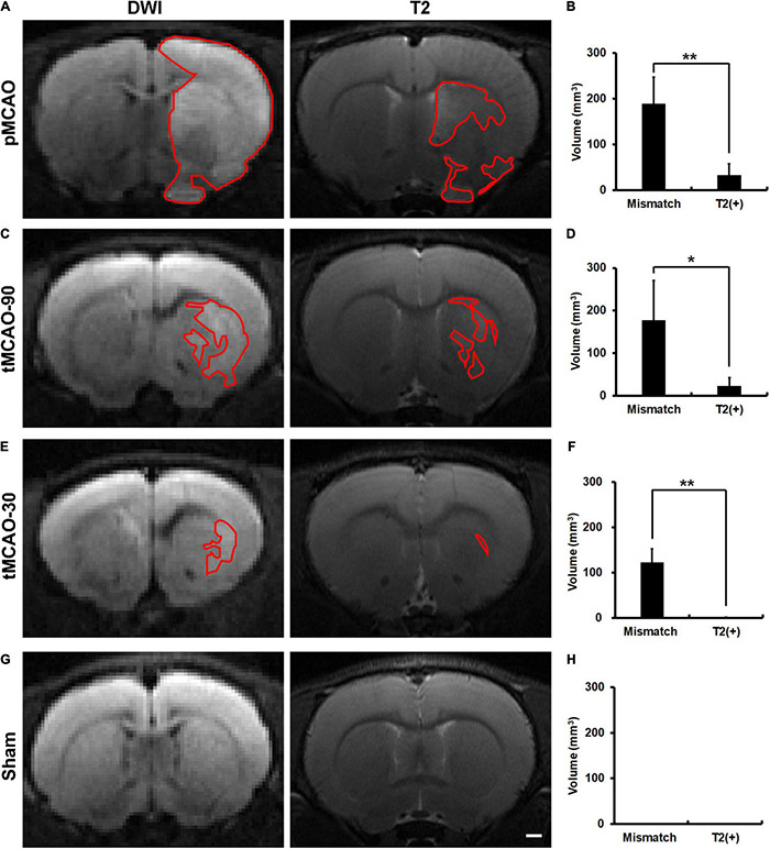
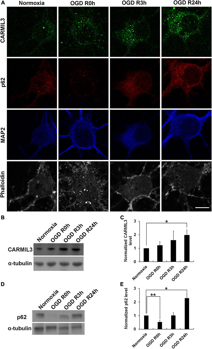
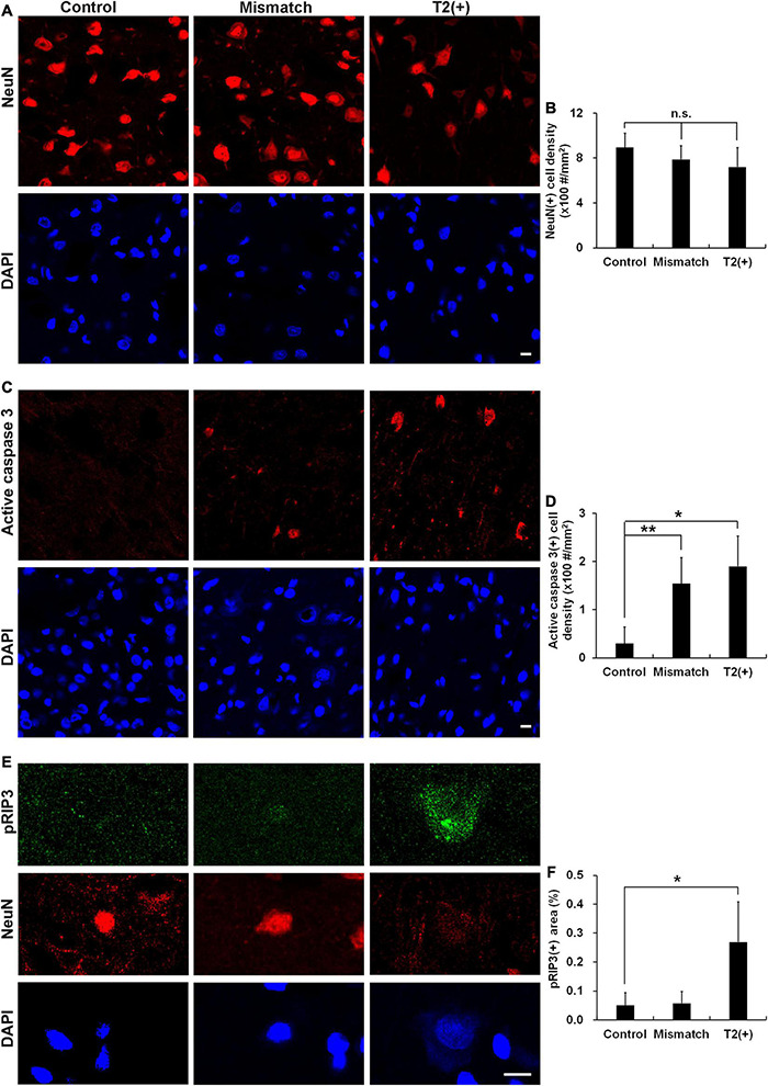
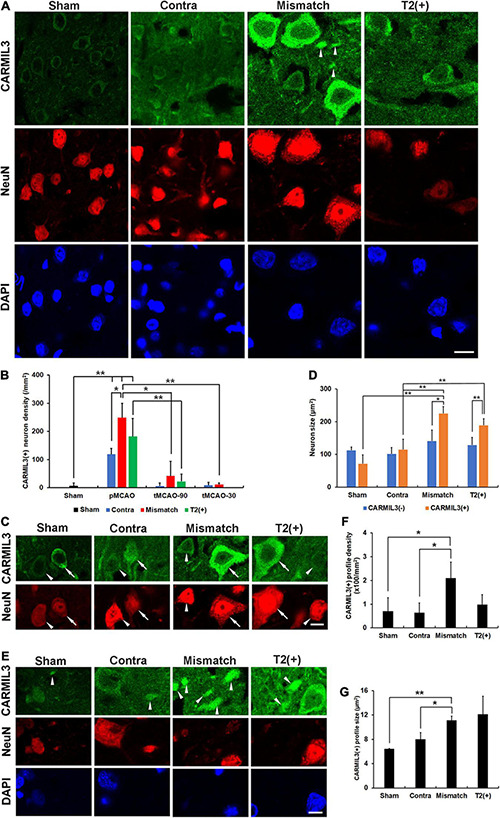
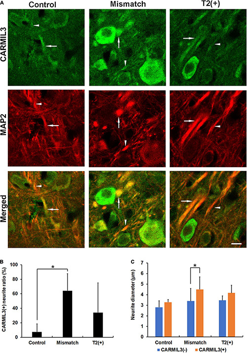
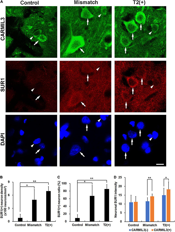
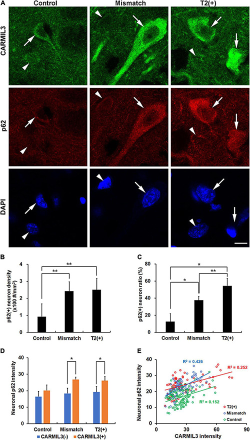
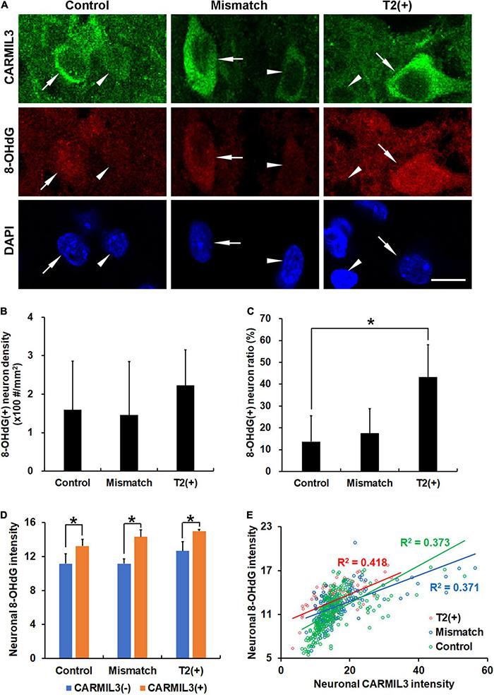
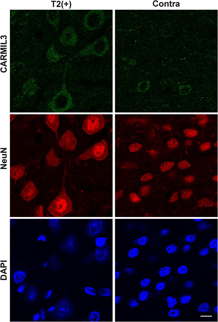
Ischemic stroke with a mismatch between diffusion-weighted imaging (DWI) and fluid-attenuated inversion recovery (FLAIR) or T2-weighted images indicates onset within 4.5 h, but the pathological substrates in the DWI-T2 mismatch and T2(+) areas remain elusive. In this study, proteomics was used to explore (1) the protein expression profiles in the T2(+), mismatch, and contralateral areas, and (2) the protein with the highest expression in the T2(+) area in the brains of male Sprague-Dawley rats within 4.5 h after middle cerebral artery occlusion (MCAO). The expression of the candidate protein was further validated in (1) rat brain subjected to MCAO, (2) rat primary cortical neuronal culture with oxygen-glucose deprivation (OGD), and (3) infarcted human brain tissues. This study showed that apoptosis was observed in the T2(+) and mismatch regions and necroptosis in the T2(+) region of rat brains after MCAO. We identified capping protein regulator and myosin 1 linker 3 (CARMIL3) as the candidate molecule in the T2(+) and mismatch areas, exclusively in neurons, predominantly in the cytoplasm, and most abundant in the mismatch area. The CARMIL3(+) neurons and neurites in the mismatch and T2(+) areas were larger than those in the control area, and associated with (1) increased expression of sulfonylurea receptor 1 (SUR1), indicating edema, (2) accumulation of p62, indicating impaired autophagy, and (3) increase in 8-hydroxy-2'-deoxyguanosine (8-OHdG), indicating oxidative stress. The increased expression of CARMIL3 was validated in a cell model of cortical neurons after OGD and in infarcted human brain tissues. In conclusion, this study shows that the mismatch and T2(+) areas within 4.5 h after ischemia are characterized by upregulated expression of CARMIL3 in neurons, particularly the mismatch area, which is associated with neuronal edema, impaired autophagy, and oxidative stress, indicating that CARMIL3 serves as a molecular signature of brain ischemia.
弥散加权成像(DWI)与液体衰减反转恢复序列(FLAIR)或T2加权图像不匹配的缺血性卒中提示发病在4.5小时内,但DWI-T2不匹配区域和T2(+)区域的病理底物仍不清楚。在本研究中,蛋白质组学被用于探索:(1)T2(+)、不匹配区域及对侧区域的蛋白质表达谱;(2)大脑中动脉闭塞(MCAO)后4.5小时内雄性Sprague-Dawley大鼠大脑T2(+)区域中表达最高的蛋白质。候选蛋白的表达在以下模型中进一步得到验证:(1)MCAO大鼠脑;(2)氧糖剥夺(OGD)的大鼠原代皮质神经元培养物;(3)梗死的人脑组织。本研究表明,MCAO后大鼠脑的T2(+)和不匹配区域出现凋亡,T2(+)区域出现坏死性凋亡。我们确定帽蛋白调节因子和肌球蛋白1连接蛋白3(CARMIL3)为T2(+)和不匹配区域的候选分子,仅在神经元中表达,主要位于细胞质中,在不匹配区域含量最丰富。不匹配区域和T2(+)区域的CARMIL3(+)神经元和神经突比对照区域的更大,并且与以下情况相关:(1)磺脲类受体1(SUR1)表达增加,提示水肿;(2)p62积累,提示自噬受损;(3)8-羟基-2'-脱氧鸟苷(8-OHdG)增加,提示氧化应激。CARMIL3表达增加在OGD后的皮质神经元细胞模型和梗死的人脑组织中得到验证。总之,本研究表明,缺血后4.5小时内的不匹配区域和T2(+)区域的特征是神经元中CARMIL3表达上调,尤其是不匹配区域,这与神经元水肿、自噬受损和氧化应激相关,表明CARMIL3是脑缺血的分子标志。