Department of Diagnostic Radiology, Kaohsiung Chang Gung Memorial Hospital, Chang Gung University College of Medicine, No. 123 Ta-Pei Road, Niao-Sung Dist, Kaohsiung City, 83305, Taiwan.
Department of Medical Imaging and Intervention, Keelung Chang Gung Memorial Hospital, Chang Gung University College of Medicine, No. 222, Maijin Road, Anle Dist, Keelung City, 204201, Taiwan.
BMC Geriatr. 2022 Jan 3;22(1):3. doi: 10.1186/s12877-021-02626-8.
The coexistence of sarcopenia and dementia in aging populations is not uncommon, and they may share common risk factors and pathophysiological pathways. This study aimed to evaluate the relationship between brain atrophy and low lean mass in the elderly with impaired cognitive function.
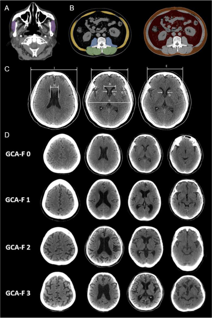
This cross-sectional study included 168 elderly patients who visited the multi-disciplinary dementia outpatient clinic at Kaohsiung Chang Gung Memorial Hospital for memory issues, between 2017 and 2019. The body composition was assessed by dual energy X-ray absorptiometry (DEXA) and CT based skeletal muscle index including L3 skeletal muscle index (L3SMI) and masseter muscle mass index (MSMI). The brain atrophy assessment was measured by CT based visual rating scale. Possible predictors of low lean mass in the elderly with cognitive impairement were identified by binary logistic regression. ROC curves were generated from binary logistic regression.
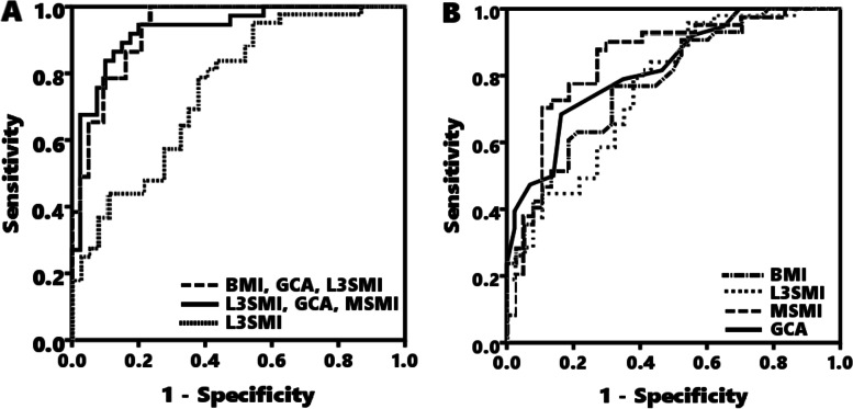
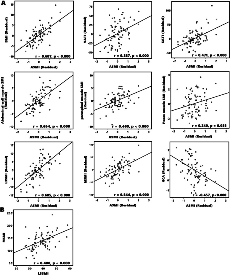
Among the 81 participants, 43 (53%) remained at a normal appendicular skeletal muscle index (ASMI), whereas 38 (47%) showed low ASMI. Compared with the normal ASMI group, subjects with low ASMI exhibited significantly lower BMI, L3SMI, and MSMI (all p < 0.05), and showed significant brain atrophy as assessed by visual rating scale (p < 0.001). The accuracy of predictive models for low ASMI in the elderly with cognitive impairment were 0.875, (Area under curve (AUC) = 0.926, 95% confidence interval [CI] 0.844-0.972) in model 1 (combination of BMI, GCA and L3SMI) and 0.885, (Area under curve (AUC) = 0.931, [CI] 0.857-0.979) in model 2 (combination of BMI, GCA and MSMI).
Global cortical atrophy and body mass index combined with either L3 skeletal muscle index or masseter skeletal muscle index can predict low lean mass in the elderly with cognitive impairment.
在老龄化人口中,肌少症和痴呆并存并不罕见,它们可能具有共同的危险因素和病理生理途径。本研究旨在评估认知功能受损的老年人中脑萎缩与低瘦体重之间的关系。
本横断面研究纳入了 2017 年至 2019 年间因记忆力问题到高雄长庚纪念医院多学科痴呆门诊就诊的 168 名老年患者。采用双能 X 射线吸收法(DEXA)和基于 CT 的骨骼肌指数(包括 L3 骨骼肌指数(L3SMI)和咬肌肌肉质量指数(MSMI))评估身体成分。采用 CT 基于视觉评分量表评估脑萎缩。通过二元逻辑回归确定认知障碍老年人低瘦体重的可能预测因素。从二元逻辑回归中生成 ROC 曲线。
在 81 名参与者中,43 名(53%)的四肢骨骼肌指数(ASMI)正常,38 名(47%)的 ASMI 较低。与 ASMI 正常组相比,低 ASMI 组的 BMI、L3SMI 和 MSMI 明显较低(均 P<0.05),并且视觉评分量表评估的脑萎缩明显(P<0.001)。用于预测认知障碍老年人低 ASMI 的预测模型的准确性在模型 1(BMI、GCA 和 L3SMI 的组合)中为 0.875(曲线下面积(AUC)=0.926,95%置信区间 [CI] 0.844-0.972)和模型 2(BMI、GCA 和 MSMI 的组合)中为 0.885(AUC=0.931,[CI] 0.857-0.979)。
总体皮质萎缩和体重指数结合 L3 骨骼肌指数或咬肌骨骼肌指数可预测认知障碍老年人的低瘦体重。