• 文献检索
  • 文档翻译
  • 深度研究
  • 学术资讯
  • Suppr Zotero 插件Zotero 插件
  • 邀请有礼
  • 套餐&价格
  • 历史记录
应用&插件
Suppr Zotero 插件Zotero 插件浏览器插件Mac 客户端Windows 客户端微信小程序
定价
高级版会员购买积分包购买API积分包
服务
文献检索文档翻译深度研究API 文档MCP 服务
关于我们
关于 Suppr公司介绍联系我们用户协议隐私条款
关注我们

Suppr 超能文献

核心技术专利:CN118964589B侵权必究
粤ICP备2023148730 号-1Suppr @ 2026

文献检索

告别复杂PubMed语法,用中文像聊天一样搜索,搜遍4000万医学文献。AI智能推荐,让科研检索更轻松。

立即免费搜索

文件翻译

保留排版,准确专业,支持PDF/Word/PPT等文件格式,支持 12+语言互译。

免费翻译文档

深度研究

AI帮你快速写综述,25分钟生成高质量综述,智能提取关键信息,辅助科研写作。

立即免费体验

六种视觉评分量表作为监测帕金森病脑萎缩体积的生物标志物

Six Visual Rating Scales as A Biomarker for Monitoring Atrophied Brain Volume in Parkinson's Disease.

作者信息

Lin Yu, Fu Ying, Zeng Yi-Fang, Hu Jian-Ping, Lin Xiao-Zhen, Cai Nai-Qing, Weng Qiang, Zhao Yi-Jing, Lin Yi, Cao Dai-Rong, Wang Ning

机构信息

1Department of Neurology and Institute of Neurology, The First Affiliated Hospital, Fujian Medical University, Fuzhou 350005, China.

2Central Laboratory, The First Affiliated Hospital, Fujian Medical University, Fuzhou 350005, China.

出版信息

Aging Dis. 2020 Oct 1;11(5):1082-1090. doi: 10.14336/AD.2019.1103. eCollection 2020 Oct.

DOI:10.14336/AD.2019.1103
PMID:33014524
原文链接:https://pmc.ncbi.nlm.nih.gov/articles/PMC7505277/
Abstract

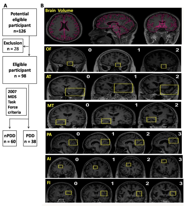
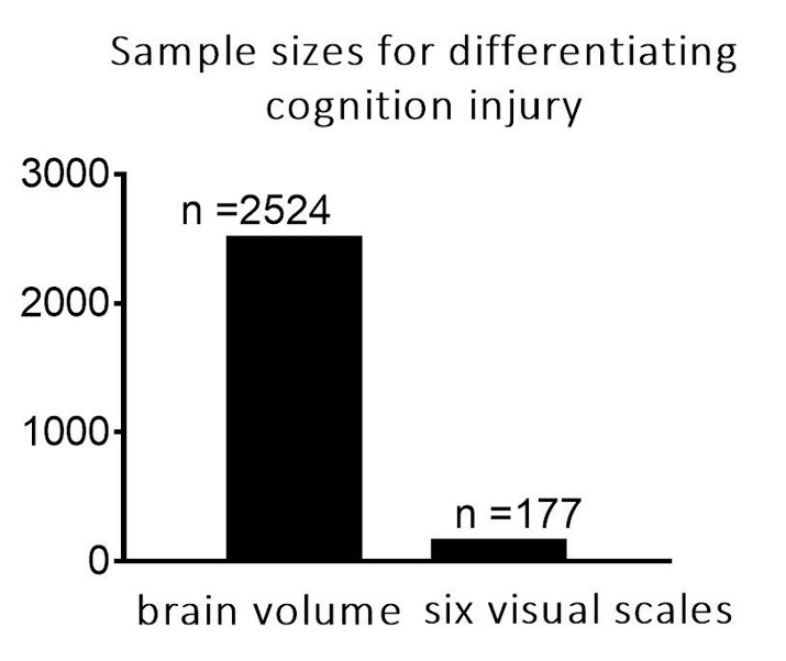
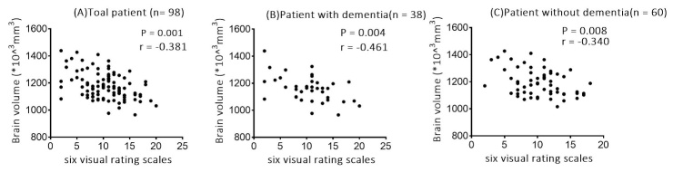
The focus of our investigation was to determine the feasibility of using six visual rating scales as whole-brain imaging markers for monitoring atrophied brain volume in Parkinson's disease (PD). This was a prospective cross-sectional single-center observational study. A total of 98 PD patients were enrolled and underwent an MRI scan and a battery of neuropsychological evaluations. The brain volume was calculated using the online resource MRICloud. Brain atrophy was rated based on six visual rating scales. Correlation analysis was performed between visual rating scores and brain volume and clinical features. We found a significant negative correlation between the total scores of visual rating scores and quantitative brain volume, indicating that six visual rating scales reliably reflect whole brain atrophy in PD. Multiple linear regression-based analyses indicated severer non-motor symptoms were significantly associated with higher scores on the visual rating scales. Furthermore, we performed sample size calculations to evaluate the superiority of visual rating scales; the result show that using total scores of visual rating scales as an outcome measure, sample sizes for differentiating cognition injury require significantly fewer subjects (n = 177) compared with using total brain volume (n = 2524). Our data support the use of the total visual rating scores rather than quantitative brain volume as a biomarker for monitoring cerebral atrophy.

摘要

我们研究的重点是确定使用六种视觉评分量表作为全脑成像标记物来监测帕金森病(PD)脑萎缩体积的可行性。这是一项前瞻性横断面单中心观察性研究。共纳入98例PD患者,他们接受了MRI扫描和一系列神经心理学评估。使用在线资源MRICloud计算脑体积。基于六种视觉评分量表对脑萎缩进行评分。对视觉评分与脑体积及临床特征进行相关性分析。我们发现视觉评分总分与定量脑体积之间存在显著负相关,表明六种视觉评分量表能可靠地反映PD患者的全脑萎缩情况。基于多元线性回归的分析表明,较严重的非运动症状与视觉评分量表上的较高分数显著相关。此外,我们进行了样本量计算以评估视觉评分量表的优越性;结果显示,将视觉评分量表总分作为结果指标时,与使用全脑体积相比,区分认知损伤所需的样本量显著更少(n = 177),而使用全脑体积时需要2524例受试者(n = 2524)。我们的数据支持使用视觉评分总分而非定量脑体积作为监测脑萎缩的生物标志物。

https://cdn.ncbi.nlm.nih.gov/pmc/blobs/da28/7505277/eb75b472a806/ad-11-5-1082-g3.jpg
https://cdn.ncbi.nlm.nih.gov/pmc/blobs/da28/7505277/30876a4eb620/ad-11-5-1082-g1.jpg
https://cdn.ncbi.nlm.nih.gov/pmc/blobs/da28/7505277/e9d3cda98eab/ad-11-5-1082-g2.jpg
https://cdn.ncbi.nlm.nih.gov/pmc/blobs/da28/7505277/eb75b472a806/ad-11-5-1082-g3.jpg
https://cdn.ncbi.nlm.nih.gov/pmc/blobs/da28/7505277/30876a4eb620/ad-11-5-1082-g1.jpg
https://cdn.ncbi.nlm.nih.gov/pmc/blobs/da28/7505277/e9d3cda98eab/ad-11-5-1082-g2.jpg
https://cdn.ncbi.nlm.nih.gov/pmc/blobs/da28/7505277/eb75b472a806/ad-11-5-1082-g3.jpg

相似文献

1
Six Visual Rating Scales as A Biomarker for Monitoring Atrophied Brain Volume in Parkinson's Disease.六种视觉评分量表作为监测帕金森病脑萎缩体积的生物标志物
Aging Dis. 2020 Oct 1;11(5):1082-1090. doi: 10.14336/AD.2019.1103. eCollection 2020 Oct.
2
Influence of diabetes mellitus on longitudinal atrophy and cognition in Parkinson's disease.糖尿病对帕金森病纵向萎缩及认知的影响。
J Neurol Sci. 2017 Jun 15;377:122-126. doi: 10.1016/j.jns.2017.04.010. Epub 2017 Apr 11.
3
Visual rating of the hippocampus in non-demented elders: Does it measure hippocampal atrophy or other indices of brain atrophy? The SMART-MR study.非痴呆老年人中海马的视觉评估:它测量的是海马萎缩还是其他脑萎缩指标?SMART-MR 研究。
Hippocampus. 2009 Nov;19(11):1115-22. doi: 10.1002/hipo.20575.
4
Multiple Visual Rating Scales Based on Structural MRI and a Novel Prediction Model Combining Visual Rating Scales and Age Stratification in the Diagnosis of Alzheimer's Disease in the Chinese Population.基于结构磁共振成像的多种视觉评定量表及结合视觉评定量表与年龄分层的新型预测模型在中国人群阿尔茨海默病诊断中的应用
Front Neurol. 2019 Feb 20;10:93. doi: 10.3389/fneur.2019.00093. eCollection 2019.
5
Visual rating of age-related white matter changes on magnetic resonance imaging: scale comparison, interrater agreement, and correlations with quantitative measurements.磁共振成像上年龄相关性白质变化的视觉评分:量表比较、评分者间一致性以及与定量测量的相关性
Stroke. 2003 Feb;34(2):441-5. doi: 10.1161/01.str.0000049766.26453.e9.
6
Grey matter atrophy in cognitively impaired Parkinson's disease.认知障碍型帕金森病患者的脑灰质萎缩。
J Neurol Neurosurg Psychiatry. 2012 Feb;83(2):188-94. doi: 10.1136/jnnp-2011-300828. Epub 2011 Sep 2.
7
Automatically computed rating scales from MRI for patients with cognitive disorders.用于认知障碍患者的 MRI 自动计算评分量表。
Eur Radiol. 2019 Sep;29(9):4937-4947. doi: 10.1007/s00330-019-06067-1. Epub 2019 Feb 22.
8
Quantitative Validation of a Visual Rating Scale for Defining High-Iron Putamen in Patients With Multiple System Atrophy.用于定义多系统萎缩患者高铁含量壳核的视觉评分量表的定量验证
Front Neurol. 2019 Sep 20;10:1014. doi: 10.3389/fneur.2019.01014. eCollection 2019.
9
A comparative study: visual rating scores and the voxel-based specific regional analysis system for Alzheimer's disease on magnetic resonance imaging among subjects with Alzheimer's disease, mild cognitive impairment, and normal cognition.一项对比研究:阿尔茨海默病患者、轻度认知障碍患者和认知正常者的磁共振成像上基于体素的特定区域分析系统与视觉评分对阿尔茨海默病的评估。
Psychogeriatrics. 2019 Mar;19(2):95-104. doi: 10.1111/psyg.12370. Epub 2018 Oct 1.
10
Depression rating scales in Parkinson's disease: critique and recommendations.帕金森病的抑郁评定量表:批判与建议
Mov Disord. 2007 Jun 15;22(8):1077-92. doi: 10.1002/mds.21333.

引用本文的文献

1
Concordance Between Amyloid-PET Quantification and Real-World Visual Reads.淀粉样蛋白PET定量与实际视觉读数之间的一致性
JAMA Neurol. 2025 Jul 28. doi: 10.1001/jamaneurol.2025.2218.
2
Predictor model for mild cognitive impairment in Parkinson's disease using visual rating scales.使用视觉评定量表的帕金森病轻度认知障碍预测模型
Neurol Sci. 2025 Jun 17. doi: 10.1007/s10072-025-08311-7.
3
Choroid plexus enlargement contributes to motor severity via regional glymphatic dysfunction in Parkinson's disease.脉络丛增大通过帕金森病中局部类淋巴系统功能障碍导致运动严重程度增加。

本文引用的文献

1
Multiple Visual Rating Scales Based on Structural MRI and a Novel Prediction Model Combining Visual Rating Scales and Age Stratification in the Diagnosis of Alzheimer's Disease in the Chinese Population.基于结构磁共振成像的多种视觉评定量表及结合视觉评定量表与年龄分层的新型预测模型在中国人群阿尔茨海默病诊断中的应用
Front Neurol. 2019 Feb 20;10:93. doi: 10.3389/fneur.2019.00093. eCollection 2019.
2
Identifying brain changes related to cognitive aging using VBM and visual rating scales.使用 VBM 和视觉评分量表识别与认知老化相关的大脑变化。
Neuroimage Clin. 2019;22:101697. doi: 10.1016/j.nicl.2019.101697. Epub 2019 Feb 5.
3
NPJ Parkinsons Dis. 2025 May 22;11(1):134. doi: 10.1038/s41531-025-00971-8.
4
A point-based cognitive impairment scoring system for southeast Asian adults.一种针对东南亚成年人的基于点数的认知障碍评分系统。
J Prev Alzheimers Dis. 2025 Apr;12(4):100069. doi: 10.1016/j.tjpad.2025.100069. Epub 2025 Jan 24.
5
Longitudinal Changes in Regional Brain Volumes and Cognition of Professional Fighters With Traumatic Encephalopathy Syndrome.外伤性脑损伤后综合征职业拳手的脑区体积和认知的纵向变化。
Neurology. 2023 Sep 12;101(11):e1118-e1126. doi: 10.1212/WNL.0000000000207594. Epub 2023 Jun 28.
6
Brain CT can predict low lean mass in the elderly with cognitive impairment: a community-dwelling study.脑 CT 可预测认知障碍老年人群的低瘦体组织量:一项社区居住者研究。
BMC Geriatr. 2022 Jan 3;22(1):3. doi: 10.1186/s12877-021-02626-8.
7
Constructing Prediction Models for Freezing of Gait by Nomogram and Machine Learning: A Longitudinal Study.通过列线图和机器学习构建步态冻结预测模型:一项纵向研究。
Front Neurol. 2021 Dec 6;12:684044. doi: 10.3389/fneur.2021.684044. eCollection 2021.
8
Levodopa-Induced Ocular Dyskinesia in an Early-Onset Parkinson Disease Patient With GBA Mutation.早发性帕金森病伴 GBA 突变患者中左旋多巴诱导的眼部运动障碍。
Clin Neuropharmacol. 2021;44(6):201-204. doi: 10.1097/WNF.0000000000000484.
9
The influence of white matter hyperintensity on cognitive impairment in Parkinson's disease.脑白质高信号对帕金森病认知障碍的影响。
Ann Clin Transl Neurol. 2021 Sep;8(9):1917-1934. doi: 10.1002/acn3.51429. Epub 2021 Jul 26.
Distinct patterns of brain atrophy in Genetic Frontotemporal Dementia Initiative (GENFI) cohort revealed by visual rating scales.
通过视觉评分量表揭示遗传额颞叶痴呆倡议 (GENFI) 队列中不同的脑萎缩模式。
Alzheimers Res Ther. 2018 May 24;10(1):46. doi: 10.1186/s13195-018-0376-9.
4
Imaging the Etiology of Apathy, Anxiety, and Depression in Parkinson's Disease: Implication for Treatment.帕金森病中淡漠、焦虑和抑郁的病因影像学研究:对治疗的启示。
Curr Neurol Neurosci Rep. 2017 Aug 18;17(10):76. doi: 10.1007/s11910-017-0788-0.
5
Parkinson's disease as a system-level disorder.帕金森病作为一种系统层面的疾病。
NPJ Parkinsons Dis. 2016 Dec 1;2:16025. doi: 10.1038/npjparkd.2016.25. eCollection 2016.
6
Mapping the order and pattern of brain structural MRI changes using change-point analysis in premanifest Huntington's disease.利用定数分析对处于前期亨廷顿病患者的大脑结构磁共振成像变化进行排序和模式分析。
Hum Brain Mapp. 2017 Oct;38(10):5035-5050. doi: 10.1002/hbm.23713. Epub 2017 Jun 28.
7
Longitudinal whole-brain atrophy and ventricular enlargement in nondemented Parkinson's disease.非痴呆型帕金森病的纵向全脑萎缩和脑室扩大。
Neurobiol Aging. 2017 Jul;55:78-90. doi: 10.1016/j.neurobiolaging.2017.03.012. Epub 2017 Mar 16.
8
The cortical thickness correlates of clinical manifestations in the mid-stage sporadic Parkinson's disease.散发性帕金森病中期临床表现的皮质厚度相关性
Neurosci Lett. 2016 Oct 28;633:279-289. doi: 10.1016/j.neulet.2016.09.042. Epub 2016 Oct 7.
9
The nonmotor features of Parkinson's disease: pathophysiology and management advances.帕金森病的非运动症状:病理生理学与治疗进展
Curr Opin Neurol. 2016 Aug;29(4):467-73. doi: 10.1097/WCO.0000000000000348.
10
Structural Imaging and Parkinson's Disease: Moving Toward Quantitative Markers of Disease Progression.结构成像与帕金森病:迈向疾病进展的定量标志物
J Parkinsons Dis. 2016 May 27;6(3):557-67. doi: 10.3233/JPD-160824.