• 文献检索
  • 文档翻译
  • 深度研究
  • 学术资讯
  • Suppr Zotero 插件Zotero 插件
  • 邀请有礼
  • 套餐&价格
  • 历史记录
应用&插件
Suppr Zotero 插件Zotero 插件浏览器插件Mac 客户端Windows 客户端微信小程序
定价
高级版会员购买积分包购买API积分包
服务
文献检索文档翻译深度研究API 文档MCP 服务
关于我们
关于 Suppr公司介绍联系我们用户协议隐私条款
关注我们

Suppr 超能文献

核心技术专利:CN118964589B侵权必究
粤ICP备2023148730 号-1Suppr @ 2026

文献检索

告别复杂PubMed语法,用中文像聊天一样搜索,搜遍4000万医学文献。AI智能推荐,让科研检索更轻松。

立即免费搜索

文件翻译

保留排版,准确专业,支持PDF/Word/PPT等文件格式,支持 12+语言互译。

免费翻译文档

深度研究

AI帮你快速写综述,25分钟生成高质量综述,智能提取关键信息,辅助科研写作。

立即免费体验

临床验证使用原型软件进行自动软骨分割以定量志愿者膝关节软骨的方法。

Clinical validation of the use of prototype software for automatic cartilage segmentation to quantify knee cartilage in volunteers.

机构信息

Department of Radiology, The Third Hospital of Hebei Medical University, Hebei Province Biomechanical Key Laboratory of Orthopedics, Shijiazhuang, Hebei, China.

MR Collaboration, Siemens Healthineers Ltd., Shanghai, China.

出版信息

BMC Musculoskelet Disord. 2022 Jan 3;23(1):19. doi: 10.1186/s12891-021-04973-4.

DOI:10.1186/s12891-021-04973-4
PMID:34980107
原文链接:https://pmc.ncbi.nlm.nih.gov/articles/PMC8725480/
Abstract

BACKGROUND

The cartilage segmentation algorithms make it possible to accurately evaluate the morphology and degeneration of cartilage. There are some factors (location of cartilage subregions, hydrarthrosis and cartilage degeneration) that may influence the accuracy of segmentation. It is valuable to evaluate and compare the accuracy and clinical value of volume and mean T2* values generated directly from automatic knee cartilage segmentation with those from manually corrected results using prototype software.

METHOD

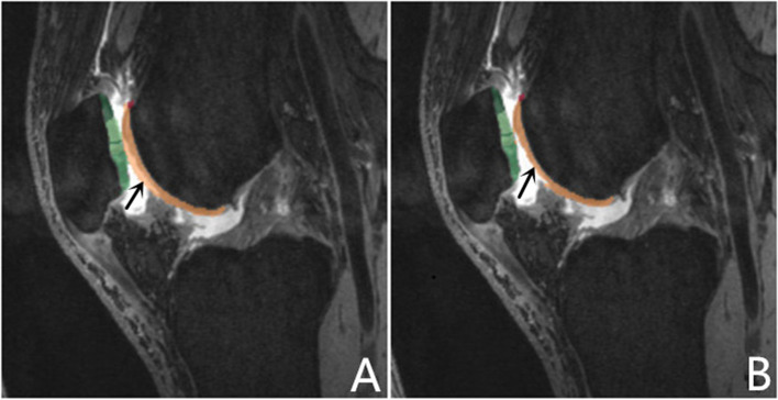
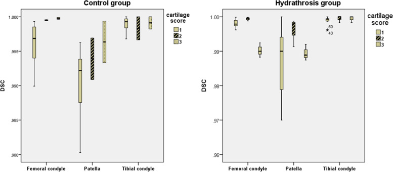
Thirty-two volunteers were recruited, all of whom underwent right knee magnetic resonance imaging examinations. Morphological images were obtained using a three-dimensional (3D) high-resolution Double-Echo in Steady-State (DESS) sequence, and biochemical images were obtained using a two-dimensional T2* mapping sequence. Cartilage score criteria ranged from 0 to 2 and were obtained using the Whole-Organ Magnetic Resonance Imaging Score (WORMS). The femoral, patellar, and tibial cartilages were automatically segmented and divided into subregions using the post-processing prototype software. Afterwards, all the subregions were carefully checked and manual corrections were done where needed. The dice coefficient correlations for each subregion by the automatic segmentation were calculated.

RESULTS

Cartilage volume after applying the manual correction was significantly lower than automatic segmentation (P < 0.05). The percentages of the cartilage volume change for each subregion after manual correction were all smaller than 5%. In all the subregions, the mean T2* relaxation time within manual corrected subregions was significantly lower than in regions after automatic segmentation (P < 0.05). The average time for the automatic segmentation of the whole knee was around 6 min, while the average time for manual correction of the whole knee was around 27 min.

CONCLUSIONS

Automatic segmentation of cartilage volume has a high dice coefficient correlation and it can provide accurate quantitative information about cartilage efficiently without individual bias. Advances in knowledge: Magnetic resonance imaging is the most promising method to detect structural changes in cartilage tissue. Unfortunately, due to the structure and morphology of the cartilages obtaining accurate segmentations can be problematic. There are some factors (location of cartilage subregions, hydrarthrosis and cartilage degeneration) that may influence segmentation accuracy. We therefore assessed the factors that influence segmentations error.

摘要

背景

软骨分割算法可实现对软骨形态和退变的精确评估。有一些因素(软骨亚区的位置、滑液和软骨退变)可能会影响分割的准确性。评估和比较直接从自动膝关节软骨分割生成的容积和平均 T2* 值与使用原型软件进行手动校正后的结果的准确性和临床价值是很有价值的。

方法

招募了 32 名志愿者,所有志愿者均接受右膝关节磁共振成像检查。形态学图像采用三维(3D)高分辨率双稳态稳态(DESS)序列获得,生化图像采用二维 T2*映射序列获得。软骨评分标准范围为 0 至 2 分,采用全器官磁共振成像评分(WORMS)获得。使用后处理原型软件对股骨、髌骨和胫骨软骨进行自动分割并划分为亚区。然后仔细检查所有亚区,并在需要时进行手动校正。通过自动分割计算每个亚区的骰子系数相关性。

结果

应用手动校正后软骨体积明显低于自动分割(P<0.05)。手动校正后各亚区软骨体积变化率均小于 5%。在所有亚区中,手动校正后的亚区平均 T2*弛豫时间均明显低于自动分割后的亚区(P<0.05)。整个膝关节的自动分割平均时间约为 6 分钟,而整个膝关节的手动校正平均时间约为 27 分钟。

结论

软骨体积的自动分割具有较高的骰子系数相关性,可在没有个体偏差的情况下高效提供软骨的准确定量信息。知识的进步:磁共振成像是检测软骨组织结构变化最有前途的方法。不幸的是,由于软骨的结构和形态,获得准确的分割可能会有问题。有一些因素(软骨亚区的位置、滑液和软骨退变)可能会影响分割的准确性。因此,我们评估了影响分割误差的因素。

https://cdn.ncbi.nlm.nih.gov/pmc/blobs/8d43/8725480/d8f0d69f4e38/12891_2021_4973_Fig2_HTML.jpg
https://cdn.ncbi.nlm.nih.gov/pmc/blobs/8d43/8725480/8b43f13d1287/12891_2021_4973_Fig1_HTML.jpg
https://cdn.ncbi.nlm.nih.gov/pmc/blobs/8d43/8725480/d8f0d69f4e38/12891_2021_4973_Fig2_HTML.jpg
https://cdn.ncbi.nlm.nih.gov/pmc/blobs/8d43/8725480/8b43f13d1287/12891_2021_4973_Fig1_HTML.jpg
https://cdn.ncbi.nlm.nih.gov/pmc/blobs/8d43/8725480/d8f0d69f4e38/12891_2021_4973_Fig2_HTML.jpg

相似文献

1
Clinical validation of the use of prototype software for automatic cartilage segmentation to quantify knee cartilage in volunteers.临床验证使用原型软件进行自动软骨分割以定量志愿者膝关节软骨的方法。
BMC Musculoskelet Disord. 2022 Jan 3;23(1):19. doi: 10.1186/s12891-021-04973-4.
2
A reproducibility study of knee cartilage volume and thickness values derived by fully automatic segmentation based on three-dimensional dual-echo in steady state data from 1.5 T and 3 T magnetic resonance imaging.基于 1.5T 和 3T 磁共振成像的三维双回波稳态数据的全自动分割得到的膝关节软骨体积和厚度值的可重复性研究。
MAGMA. 2024 Feb;37(1):69-82. doi: 10.1007/s10334-023-01122-x. Epub 2023 Oct 10.
3
Longitudinal study of the morphological and T2* changes of knee cartilages of marathon runners using prototype software for automatic cartilage segmentation.利用自动软骨分割原型软件对马拉松运动员膝关节软骨的形态和 T2*变化进行的纵向研究。
Br J Radiol. 2021 Mar 1;94(1119):20200833. doi: 10.1259/bjr.20200833. Epub 2021 Feb 5.
4
Use of 2D U-Net Convolutional Neural Networks for Automated Cartilage and Meniscus Segmentation of Knee MR Imaging Data to Determine Relaxometry and Morphometry.使用 2D U-Net 卷积神经网络对膝关节 MRI 数据进行自动软骨和半月板分割以确定弛豫度和形态测量学。
Radiology. 2018 Jul;288(1):177-185. doi: 10.1148/radiol.2018172322. Epub 2018 Mar 27.
5
Reproducibility of an Automated Quantitative MRI Assessment of Low-Grade Knee Articular Cartilage Lesions.一种用于评估低级别膝关节关节软骨病变的自动定量 MRI 评估方法的可重复性。
Cartilage. 2021 Dec;13(1_suppl):646S-657S. doi: 10.1177/1947603520961165. Epub 2020 Sep 29.
6
Interscan measurement error of knee cartilage thickness and projected cartilage area ratio at 9 regions and 45 subregions by fully automatic three-dimensional MRI analysis.全自动三维 MRI 分析在 9 个区域和 45 个亚区测量膝关节软骨厚度和投影软骨面积比的误差。
Eur J Radiol. 2021 Jun;139:109700. doi: 10.1016/j.ejrad.2021.109700. Epub 2021 Apr 8.
7
Automatic segmentation of the glenohumeral cartilages from magnetic resonance images.从磁共振图像中自动分割肩肱关节软骨。
Med Phys. 2016 Oct;43(10):5370. doi: 10.1118/1.4961011.
8
Open Source Software for Automatic Subregional Assessment of Knee Cartilage Degradation Using Quantitative T2 Relaxometry and Deep Learning.开源软件用于基于定量 T2 弛豫率和深度学习的膝关节软骨退变的自动亚区评估。
Cartilage. 2021 Dec;13(1_suppl):747S-756S. doi: 10.1177/19476035211042406. Epub 2021 Sep 8.
9
T2 mapping of articular cartilage in the healthy knee using a magnetic resonance segmentation.使用磁共振分割技术对健康膝关节的关节软骨进行T2映射。
Zhongguo Yi Xue Ke Xue Yuan Xue Bao. 2011 Feb;33(1):66-70. doi: 10.3881/j.issn.1000-503X.2011.01.014.
10
Simultaneous super-resolution and contrast synthesis of routine clinical magnetic resonance images of the knee for improving automatic segmentation of joint cartilage: data from the Osteoarthritis Initiative.膝关节常规临床磁共振图像的同步超分辨率和对比度合成以改善关节软骨自动分割:来自骨关节炎倡议组织的数据
Med Phys. 2020 Oct;47(10):4939-4948. doi: 10.1002/mp.14421. Epub 2020 Aug 15.

引用本文的文献

1
3D-DESS MRI with CAIPIRINHA two- and fourfold acceleration for quantitatively assessing knee cartilage morphology.基于 CAIPIRINHA 双次和四次加速的 3D-DESS MRI 定量评估膝关节软骨形态。
Skeletal Radiol. 2024 Aug;53(8):1481-1494. doi: 10.1007/s00256-024-04605-7. Epub 2024 Feb 13.
2
Digital health in musculoskeletal care: where are we heading?数字健康在肌肉骨骼护理中的应用:我们的发展方向在哪里?
BMC Musculoskelet Disord. 2023 Mar 14;24(1):192. doi: 10.1186/s12891-023-06309-w.
3
Analysis of Knee Joint Injury Caused by Physical Training of Freshmen Students Based on 3T MRI and Automatic Cartilage Segmentation Technology: A Prospective Study.

本文引用的文献

1
The International Workshop on Osteoarthritis Imaging Knee MRI Segmentation Challenge: A Multi-Institute Evaluation and Analysis Framework on a Standardized Dataset.骨关节炎影像学膝关节MRI分割挑战赛国际研讨会:基于标准化数据集的多机构评估与分析框架
Radiol Artif Intell. 2021 Feb 10;3(3):e200078. doi: 10.1148/ryai.2021200078. eCollection 2021 May.
2
Deep convolutional neural network and 3D deformable approach for tissue segmentation in musculoskeletal magnetic resonance imaging.基于深度卷积神经网络和 3D 可变形方法的肌肉骨骼磁共振成像中的组织分割。
Magn Reson Med. 2018 Apr;79(4):2379-2391. doi: 10.1002/mrm.26841. Epub 2017 Jul 21.
3
基于 3T MRI 和自动软骨分割技术分析新生学生体能训练所致膝关节损伤:一项前瞻性研究。
Front Endocrinol (Lausanne). 2022 May 9;13:839112. doi: 10.3389/fendo.2022.839112. eCollection 2022.
3D double-echo steady-state sequence assessment of hip joint cartilage and labrum at 3 Tesla: comparative analysis of magnetic resonance imaging and intraoperative data.
3T 下髋关节软骨和盂唇的 3D 双回波稳态序列评估:磁共振成像与术中数据的对比分析。
Eur Radiol. 2017 Oct;27(10):4360-4371. doi: 10.1007/s00330-017-4834-8. Epub 2017 Apr 21.
4
Correlation between T2* (T2 star) relaxation time and cervical intervertebral disc degeneration: An observational study.T2*(T2星)弛豫时间与颈椎间盘退变的相关性:一项观察性研究。
Medicine (Baltimore). 2016 Nov;95(47):e4502. doi: 10.1097/MD.0000000000004502.
5
The immediate effect of long-distance running on T2 and T2* relaxation times of articular cartilage of the knee in young healthy adults at 3.0 T MR imaging.3.0T磁共振成像下长跑对年轻健康成年人膝关节软骨T2及T2*弛豫时间的即时影响
Br J Radiol. 2016 Aug;89(1064):20151075. doi: 10.1259/bjr.20151075. Epub 2016 Jun 23.
6
Comparison of T1ρ and T2* Relaxation Mapping in Patients with Different Grades of Disc Degeneration at 3T MR.3T磁共振成像下不同椎间盘退变程度患者的T1ρ与T2*弛豫成像比较
Med Sci Monit. 2015 Jul 4;21:1934-41. doi: 10.12659/MSM.894406.
7
Morphological MR imaging of the articular cartilage of the knee at 3 T-comparison of standard and novel 3D sequences.3T下膝关节软骨的形态学磁共振成像——标准与新型3D序列的比较
Insights Imaging. 2015 Jun;6(3):285-93. doi: 10.1007/s13244-015-0405-1. Epub 2015 Apr 9.
8
Computer-aided diagnosis of early knee osteoarthritis based on MRI T2 mapping.基于MRI T2映射的早期膝关节骨关节炎计算机辅助诊断
Biomed Mater Eng. 2014;24(6):3379-88. doi: 10.3233/BME-141161.
9
Fully automated segmentation of cartilage from the MR images of knee using a multi-atlas and local structural analysis method.使用多图谱和局部结构分析方法从膝关节磁共振图像中全自动分割软骨。
Med Phys. 2014 Sep;41(9):092303. doi: 10.1118/1.4893533.
10
T2* mapping for articular cartilage assessment: principles, current applications, and future prospects.用于关节软骨评估的T2* 成像:原理、当前应用及未来前景
Skeletal Radiol. 2014 Oct;43(10):1429-45. doi: 10.1007/s00256-014-1852-3. Epub 2014 Mar 19.