• 文献检索
  • 文档翻译
  • 深度研究
  • 学术资讯
  • Suppr Zotero 插件Zotero 插件
  • 邀请有礼
  • 套餐&价格
  • 历史记录
应用&插件
Suppr Zotero 插件Zotero 插件浏览器插件Mac 客户端Windows 客户端微信小程序
定价
高级版会员购买积分包购买API积分包
服务
文献检索文档翻译深度研究API 文档MCP 服务
关于我们
关于 Suppr公司介绍联系我们用户协议隐私条款
关注我们

Suppr 超能文献

核心技术专利:CN118964589B侵权必究
粤ICP备2023148730 号-1Suppr @ 2026

文献检索

告别复杂PubMed语法,用中文像聊天一样搜索,搜遍4000万医学文献。AI智能推荐,让科研检索更轻松。

立即免费搜索

文件翻译

保留排版,准确专业,支持PDF/Word/PPT等文件格式,支持 12+语言互译。

免费翻译文档

深度研究

AI帮你快速写综述,25分钟生成高质量综述,智能提取关键信息,辅助科研写作。

立即免费体验

症状性轻至中度颈动脉狭窄患者斑块成分和形态的性别差异。

Sex Differences in Plaque Composition and Morphology Among Symptomatic Patients With Mild-to-Moderate Carotid Artery Stenosis.

机构信息

Department of Radiology and Nuclear Medicine (D.H.K.v.D.-N., N.C.M.v.E., K.D., A.v.d.L., D.B.), Erasmus University Medical Center Rotterdam, Rotterdam, the Netherlands.

Department of Neurology (D.H.K.v.D.-N., P.J.K.), Erasmus University Medical Center Rotterdam, Rotterdam, the Netherlands.

出版信息

Stroke. 2022 Feb;53(2):370-378. doi: 10.1161/STROKEAHA.121.036564. Epub 2022 Jan 5.

DOI:10.1161/STROKEAHA.121.036564
PMID:34983237
原文链接:https://pmc.ncbi.nlm.nih.gov/articles/PMC8785521/
Abstract

BACKGROUND AND PURPOSE

Incidence of ischemic stroke differs between men and women, with substantially higher rates in men. The underlying mechanism of this difference remains poorly understood but may be because of differences in carotid atherosclerosis. Using an in-depth imaging-based approach, we investigated differences between carotid plaque composition and morphology in male and female patients with stroke, taking into account differences in total plaque burden. Additionally, we investigated all possible within-artery combinations of plaque characteristics to explore differences between various plaque phenotypes.

METHODS

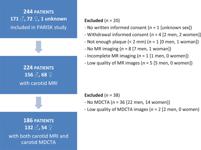
We included 156 men and 68 women from the PARISK (Plaque At Risk) study, a prospective cohort study of patients with recent ischemic cerebrovascular symptoms and <70% ipsilateral carotid stenosis. Plaque characteristics (intraplaque hemorrhage [IPH], lipid-rich necrotic core [LRNC], calcifications, thin-or-ruptured fibrous cap, ulcerations, total plaque volume) were assessed with magnetic resonance imaging and multidetector-row computed tomography angiography. We used multivariable logistic and linear regression analyses to assess sex differences in plaque characteristics.

RESULTS

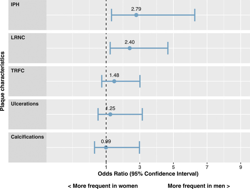
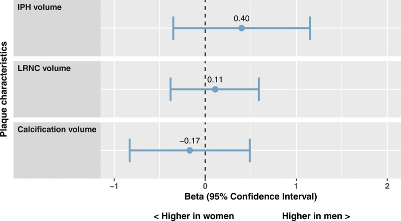
We found significant difference in total plaque volume between men and women (β=22.9 mm [95% CI, 15.4-30.5]; mean volume in men 1399±425 mm, in women 1011±242 mm). Additionally, men were more likely to have IPH (odds ratio [OR]=2.8 [95% CI, 1.3-6.3]; IPH proportion in men 49%, in women 16%) and LRNC (OR=2.4 [95% CI, 1.2-4.7]; LRNC proportion in men 73%, in women 41%) even after adjustment for total plaque volume. We found no sex-specific differences in plaque volume-corrected volumes of IPH, LRNC, and calcifications. In terms of coexistence of plaque characteristics, we found that men had more often a plaque with coexistence of calcifications, LRNC, and IPH (OR=2.7 [95% CI, 1.2-7.0]), with coexistence of thin-or-ruptured fibrous cap/ulcerations, LRNC, and IPH (OR=2.4 [95% CI, 1.1-5.9]), and with coexistence of all plaque characteristics (OR=3.0 [95% CI, 1.2-8.6]).

CONCLUSIONS

In symptomatic patients with mild-to-moderate carotid stenosis, men are more likely to have a high-risk carotid plaque with IPH and LRNC than women, regardless of total plaque burden. Men also have more often a plaque with multiple vulnerable plaque components, which could comprise an even higher stroke risk. Registration: URL: https://www.clinicaltrials.gov; Unique identifier: NCT01208025.

摘要

背景与目的

男性和女性的缺血性脑卒中发病率不同,男性发病率明显更高。这种差异的潜在机制仍知之甚少,但可能是由于颈动脉粥样硬化的差异所致。我们采用一种深入的基于影像学的方法,研究了男性和女性脑卒中患者颈动脉斑块成分和形态的差异,同时考虑了总斑块负担的差异。此外,我们还研究了动脉内所有可能的斑块特征组合,以探讨各种斑块表型之间的差异。

方法

我们纳入了来自 PARISK(斑块风险)研究的 156 名男性和 68 名女性患者,这是一项针对近期缺血性脑血管症状和同侧颈动脉狭窄<70%的患者的前瞻性队列研究。使用磁共振成像和多排螺旋 CT 血管造影评估斑块特征(斑块内出血[IPH]、富含脂质的坏死核心[LRNC]、钙化、薄或破裂的纤维帽、溃疡、总斑块体积)。我们使用多变量逻辑和线性回归分析来评估性别与斑块特征之间的差异。

结果

我们发现男性和女性之间的总斑块体积存在显著差异(β=22.9 mm [95%置信区间,15.4-30.5];男性平均体积为 1399±425 mm,女性为 1011±242 mm)。此外,即使在调整总斑块体积后,男性也更有可能出现 IPH(优势比[OR]=2.8 [95%置信区间,1.3-6.3];男性中 IPH 的比例为 49%,女性中为 16%)和 LRNC(OR=2.4 [95%置信区间,1.2-4.7];男性中 LRNC 的比例为 73%,女性中为 41%)。我们没有发现斑块体积校正后的 IPH、LRNC 和钙化体积在性别之间存在差异。就斑块特征的共存而言,我们发现男性更常出现钙化、LRNC 和 IPH 共存的斑块(OR=2.7 [95%置信区间,1.2-7.0])、LRNC 和 IPH 共存的薄或破裂纤维帽/溃疡斑块(OR=2.4 [95%置信区间,1.1-5.9]),以及所有斑块特征共存的斑块(OR=3.0 [95%置信区间,1.2-8.6])。

结论

在有轻度至中度颈动脉狭窄的症状性患者中,男性比女性更有可能出现伴有 IPH 和 LRNC 的高危颈动脉斑块,而不论总斑块负担如何。男性还更常出现具有多种易损斑块成分的斑块,这可能构成更高的卒中风险。

注册

网址:https://www.clinicaltrials.gov;唯一标识符:NCT01208025。

https://cdn.ncbi.nlm.nih.gov/pmc/blobs/0866/8820767/dd15dab0111d/str-53-370-g006.jpg
https://cdn.ncbi.nlm.nih.gov/pmc/blobs/0866/8820767/ee8f9a69d0dd/str-53-370-g001.jpg
https://cdn.ncbi.nlm.nih.gov/pmc/blobs/0866/8820767/cecb2caac4e1/str-53-370-g004.jpg
https://cdn.ncbi.nlm.nih.gov/pmc/blobs/0866/8820767/6ac178421f6a/str-53-370-g005.jpg
https://cdn.ncbi.nlm.nih.gov/pmc/blobs/0866/8820767/dd15dab0111d/str-53-370-g006.jpg

相似文献

1
Sex Differences in Plaque Composition and Morphology Among Symptomatic Patients With Mild-to-Moderate Carotid Artery Stenosis.症状性轻至中度颈动脉狭窄患者斑块成分和形态的性别差异。
Stroke. 2022 Feb;53(2):370-378. doi: 10.1161/STROKEAHA.121.036564. Epub 2022 Jan 5.
2
Size of carotid artery intraplaque hemorrhage and acute ischemic stroke: a cardiovascular magnetic resonance Chinese atherosclerosis risk evaluation study.颈动脉斑块内出血与急性缺血性脑卒中的大小:一项心血管磁共振中国动脉粥样硬化风险评估研究。
J Cardiovasc Magn Reson. 2019 Jul 1;21(1):36. doi: 10.1186/s12968-019-0548-1.
3
Contemporary carotid imaging: from degree of stenosis to plaque vulnerability.当代颈动脉成像:从狭窄程度到斑块易损性
J Neurosurg. 2016 Jan;124(1):27-42. doi: 10.3171/2015.1.JNS142452. Epub 2015 Jul 31.
4
Carotid Plaque Characteristics Predict Recurrent Ischemic Stroke and TIA: The PARISK (Plaque At RISK) Study.颈动脉斑块特征预测复发性缺血性中风和短暂性脑缺血发作:PARISK(风险斑块)研究。
JACC Cardiovasc Imaging. 2022 Oct;15(10):1715-1726. doi: 10.1016/j.jcmg.2022.04.003. Epub 2022 Jun 15.
5
Association between fibrinogen and fibrinogen γ' and atherosclerotic plaque morphology and composition in symptomatic carotid artery stenosis: Plaque-At-RISK study.纤维蛋白原和纤维蛋白原 γ'与症状性颈动脉狭窄动脉粥样硬化斑块形态和成分的关系:Plaque-At-RISK 研究。
Thromb Res. 2019 May;177:130-135. doi: 10.1016/j.thromres.2019.02.030. Epub 2019 Feb 27.
6
Intraplaque hemorrhage, fibrous cap status, and microembolic signals in symptomatic patients with mild to moderate carotid artery stenosis: the Plaque at RISK study.症状性轻至中度颈动脉狭窄患者的斑块内出血、纤维帽状态及微栓塞信号:高危斑块研究
Stroke. 2014 Nov;45(11):3423-6. doi: 10.1161/STROKEAHA.114.006800. Epub 2014 Sep 25.
7
MR imaging of vulnerable carotid plaque.易损性颈动脉斑块的磁共振成像
Cardiovasc Diagn Ther. 2020 Aug;10(4):1019-1031. doi: 10.21037/cdt.2020.03.12.
8
Comparison of Carotid Plaque Characteristics Between Men and Women Using Magnetic Resonance Vessel Wall Imaging: A Chinese Atherosclerosis Risk Evaluation Study.基于磁共振血管壁成像的中国动脉粥样硬化风险评估研究:男性与女性颈动脉斑块特征的比较。
J Magn Reson Imaging. 2021 Aug;54(2):646-654. doi: 10.1002/jmri.27576. Epub 2021 Feb 27.
9
Plaque vulnerability in patients with high- and moderate-grade carotid stenosis - comparison of plaque features on MRI with histopathological findings.颈动脉中重度狭窄患者斑块易损性——MRI 斑块特征与组织病理学发现的比较。
Swiss Med Wkly. 2020 Feb 17;150:w20174. doi: 10.4414/smw.2020.20174. eCollection 2020 Feb 10.
10
Lipoprotein(a) levels and atherosclerotic plaque characteristics in the carotid artery: The Plaque at RISK (PARISK) study.脂蛋白(a)水平与颈动脉粥样硬化斑块特征:风险斑块研究(PARISK)。
Atherosclerosis. 2021 Jul;329:22-29. doi: 10.1016/j.atherosclerosis.2021.06.004. Epub 2021 Jun 11.

引用本文的文献

1
Development and validation of a machine learning model for predicting vulnerable carotid plaques using routine blood biomarkers and derived indicators: insights into sex-related risk patterns.使用常规血液生物标志物和衍生指标预测易损颈动脉斑块的机器学习模型的开发与验证:对性别相关风险模式的见解
Cardiovasc Diabetol. 2025 Aug 10;24(1):326. doi: 10.1186/s12933-025-02867-6.
2
Impaired dynamic cerebral autoregulation predicts silent ischemic lesions after carotid artery stenting: A hemodynamic biomarker beyond plaque morphology.动态脑自动调节功能受损可预测颈动脉支架置入术后的无症状缺血性病变:一种超越斑块形态的血流动力学生物标志物。
J Cereb Blood Flow Metab. 2025 Jul 31:271678X251362000. doi: 10.1177/0271678X251362000.
3

本文引用的文献

1
Prediction of Stroke Risk by Detection of Hemorrhage in Carotid Plaques: Meta-Analysis of Individual Patient Data.基于颈动脉斑块出血检测预测卒中风险:个体患者数据分析的荟萃分析。
JACC Cardiovasc Imaging. 2020 Feb;13(2 Pt 1):395-406. doi: 10.1016/j.jcmg.2019.03.028. Epub 2019 Jun 12.
2
Imaging biomarkers of vulnerable carotid plaques for stroke risk prediction and their potential clinical implications.易损颈动脉斑块的影像学生物标志物用于卒中风险预测及其潜在临床意义。
Lancet Neurol. 2019 Jun;18(6):559-572. doi: 10.1016/S1474-4422(19)30035-3. Epub 2019 Apr 4.
3
User's guide to correlation coefficients.
Regressing calcifications in the carotid artery: What's going on? A case series.
颈动脉钙化的消退:怎么回事?病例系列
Radiol Case Rep. 2025 Jul 4;20(9):4797-4801. doi: 10.1016/j.radcr.2025.05.108. eCollection 2025 Sep.
4
Association of Carotid Plaque Calcification Attenuation With Intraplaque Hemorrhage Volume: 3D-Segmentation Analysis.颈动脉斑块钙化衰减与斑块内出血体积的关联:三维分割分析
J Neuroimaging. 2025 Jul-Aug;35(4):e70071. doi: 10.1111/jon.70071.
5
Sex differences in features of atherosclerotic plaques as revealed by various imaging techniques: historical review.各种成像技术揭示的动脉粥样硬化斑块特征中的性别差异:历史回顾
Front Physiol. 2025 May 26;16:1579885. doi: 10.3389/fphys.2025.1579885. eCollection 2025.
6
In-Depth Carotid Calcification Morphometrics and Their Temporal Changes Are Associated with Cardiovascular Risk Factors in Patients with Recent Ischemic Event: The Plaque At Risk Study.深度颈动脉钙化形态计量学及其时间变化与近期缺血性事件患者的心血管危险因素相关:危险斑块研究
Cerebrovasc Dis. 2025 May 2:1-11. doi: 10.1159/000546164.
7
Carotid artery segmentation in computed tomography angiography (CTA) using multi-scale deep supervision with Swin-UNet and advanced data augmentation.使用带有Swin-UNet的多尺度深度监督和先进数据增强技术在计算机断层扫描血管造影(CTA)中进行颈动脉分割
Quant Imaging Med Surg. 2025 Apr 1;15(4):3161-3175. doi: 10.21037/qims-24-2087. Epub 2025 Mar 28.
8
Biomechanical assessment of vulnerable plaque: from histological evidence to ultrasound elastography and image-based computational patient-specific modelling.易损斑块的生物力学评估:从组织学证据到超声弹性成像及基于图像的个体化计算模型
Front Bioeng Biotechnol. 2025 Mar 7;13:1478408. doi: 10.3389/fbioe.2025.1478408. eCollection 2025.
9
Unravelling the role of carotid atherosclerosis in predicting cardiovascular disease risk: A review.解读颈动脉粥样硬化在预测心血管疾病风险中的作用:综述
ARYA Atheroscler. 2024;20(5):52-59. doi: 10.48305/arya.2024.41271.2862.
10
Prevalence and associated risk factors of carotid plaque and artery stenosis in China: a population-based study.中国颈动脉斑块和动脉狭窄的患病率及相关危险因素:一项基于人群的研究。
Front Med. 2025 Feb;19(1):64-78. doi: 10.1007/s11684-024-1088-0. Epub 2024 Nov 27.
相关系数用户指南。
Turk J Emerg Med. 2018 Aug 7;18(3):91-93. doi: 10.1016/j.tjem.2018.08.001. eCollection 2018 Sep.
4
No Association between Thrombin Generation and Intra-Plaque Haemorrhage in Symptomatic Carotid Atherosclerotic Plaques: The Plaque at RISK (PARISK) Study.症状性颈动脉粥样硬化斑块中凝血酶生成与斑块内出血之间无关联:斑块风险研究(PARISK)。
Thromb Haemost. 2018 Aug;118(8):1461-1469. doi: 10.1055/s-0038-1666858. Epub 2018 Jul 4.
5
Determinants of carotid atherosclerotic plaque burden in a stroke-free population.无卒中人群中颈动脉粥样硬化斑块负荷的决定因素。
Atherosclerosis. 2016 Dec;255:186-192. doi: 10.1016/j.atherosclerosis.2016.10.030. Epub 2016 Oct 14.
6
Optimal Prediction of Carotid Intraplaque Hemorrhage Using Clinical and Lumen Imaging Markers.利用临床和管腔成像标志物对颈动脉斑块内出血进行最佳预测
AJNR Am J Neuroradiol. 2015 Dec;36(12):2360-6. doi: 10.3174/ajnr.A4454. Epub 2015 Sep 3.
7
Intraplaque Hemorrhage and the Plaque Surface in Carotid Atherosclerosis: The Plaque At RISK Study (PARISK).颈动脉粥样硬化中的斑块内出血与斑块表面:斑块风险研究(PARISK)
AJNR Am J Neuroradiol. 2015 Nov;36(11):2127-33. doi: 10.3174/ajnr.A4414. Epub 2015 Aug 6.
8
Plaque At RISK (PARISK): prospective multicenter study to improve diagnosis of high-risk carotid plaques.易损斑块研究(PARISK):一项旨在改善高危颈动脉斑块诊断的前瞻性多中心研究。
Int J Stroke. 2014 Aug;9(6):747-54. doi: 10.1111/ijs.12167. Epub 2013 Oct 21.
9
Carotid plaque MRI and stroke risk: a systematic review and meta-analysis.颈动脉斑块 MRI 与卒中风险:系统评价和荟萃分析。
Stroke. 2013 Nov;44(11):3071-7. doi: 10.1161/STROKEAHA.113.002551. Epub 2013 Aug 29.
10
Sex differences of high-risk carotid atherosclerotic plaque with less than 50% stenosis in asymptomatic patients: an in vivo 3T MRI study.无症状患者中狭窄程度小于 50%的高危颈动脉粥样硬化斑块的性别差异:一项体内 3T MRI 研究。
AJNR Am J Neuroradiol. 2013 May;34(5):1049-55, S1. doi: 10.3174/ajnr.A3399. Epub 2012 Nov 29.