Proteomics Department, Aravind Medical Research Foundation, Dr. G. Venkataswamy Eye Research Institute, Aravind Eye Care System, Madurai, Tamil Nadu, India.
Department of Biotechnology, Alagappa University, Karaikudi, Tamil Nadu, India.
BMC Genomics. 2022 Jan 5;23(1):5. doi: 10.1186/s12864-021-08218-5.
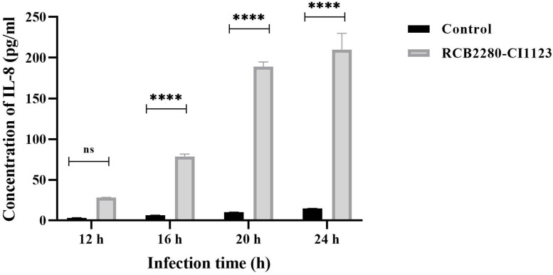
Aspergillus flavus, one of the causative agents of human fungal keratitis, can be phagocytosed by human corneal epithelial (HCE) cells and the conidia containing phagosomes mature into phagolysosomes. But the immunological responses of human corneal epithelial cells interacting with A. flavus are not clear. In this study, we report the expression of immune response related genes of HCE cells exposed to A. flavus spores using targeted transcriptomics.
Human corneal epithelial cell line and primary cultures were grown in a six-well plate and used for coculture experiments. Internalization of the conidia was confirmed by immunofluorescence microscopy of the colocalized endosomal markers CD71 and LAMP1. Total RNA was isolated, and the quantity and quality of the isolated RNA were assessed using Qubit and Bioanalyzer. NanoString nCounter platform was used for the analysis of mRNA abundance using the Human Immunology panel. R-package and nSolver software were used for data analysis. KEGG and FunRich 3.1.3 tools were used to analyze the differentially expressed genes.
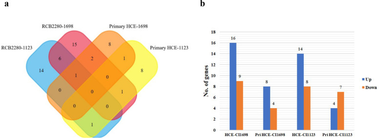
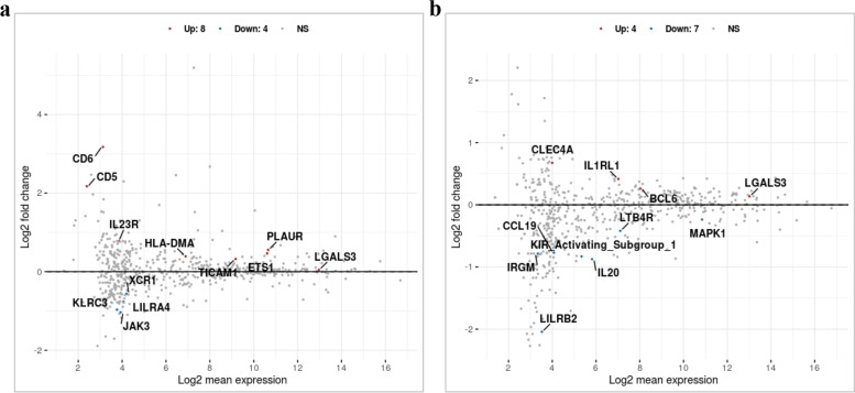
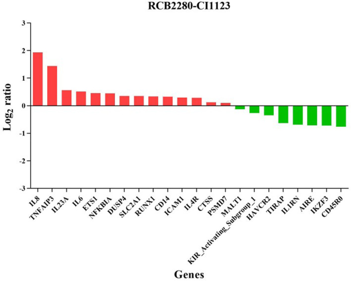
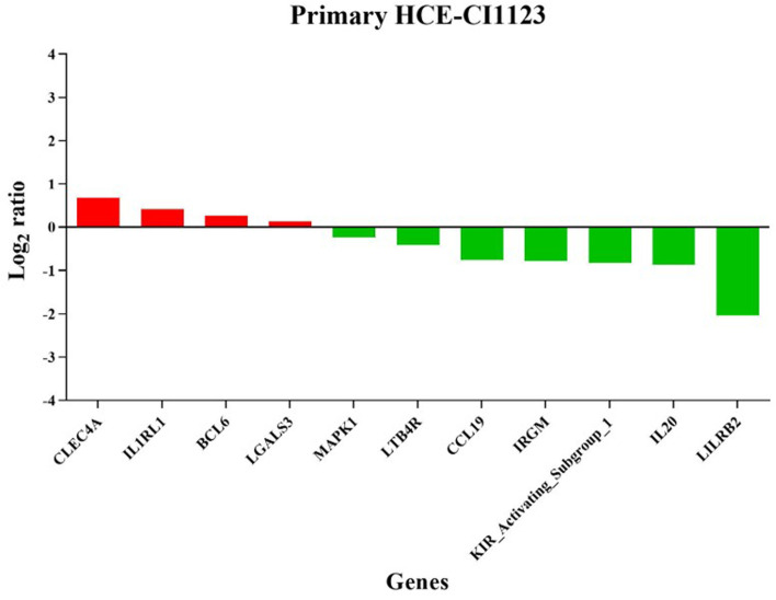
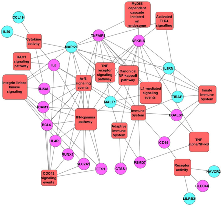
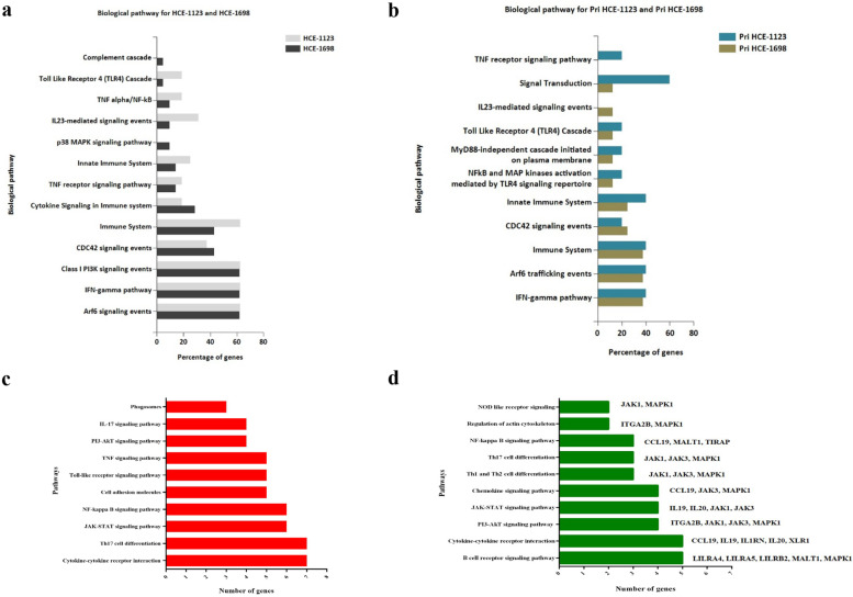
Different morphotypes of conidia were observed after 6 h of coculture with human corneal epithelial cells and found to be internalized by epithelial cells. NanoString profiling showed more than 20 differentially expressed genes in immortalized human corneal epithelial cell line and more than ten differentially expressed genes in primary corneal epithelial cells. Distinct set of genes were altered in their expression in cell line and primary corneal epithelial cells. KEGG pathway analysis revealed that genes associated with TNF signaling, NF-KB signaling, and Th17 signaling were up-regulated, and genes associated with chemokine signaling and B cell receptor signaling were down regulated. FunRich pathway analysis showed that pathways such as CDC42 signaling, PI3K signaling, and Arf6 trafficking events were activated by the clinical isolates CI1123 and CI1698 in both type of cells.
Combining the transcript analysis data from cell lines and primary cultures, we showed the up regulation of immune defense genes in A. flavus infected cells. At the same time, chemokine signaling and B cell signaling pathways are downregulated. The variability in the expression levels in the immortalized cell line and the primary cultures is likely due to the variable epigenetic reprogramming in the immortalized cells and primary cultures in the absence of any changes in the genome. It highlights the importance of using both cell types in host-pathogen interaction studies.
黄曲霉菌是人类真菌角膜炎的病原体之一,可被人角膜上皮(HCE)细胞吞噬,含孢子的吞噬体成熟为吞噬溶酶体。但是,人角膜上皮细胞与黄曲霉菌相互作用的免疫反应尚不清楚。在这项研究中,我们通过靶向转录组学报告了暴露于黄曲霉菌孢子的 HCE 细胞的免疫反应相关基因的表达。
人角膜上皮细胞系和原代培养物在六孔板中生长,并用于共培养实验。通过共定位内体标记物 CD71 和 LAMP1 的免疫荧光显微镜观察孢子的内化。使用 Qubit 和 Bioanalyzer 评估分离 RNA 的数量和质量。使用 NanoString nCounter 平台分析使用人类免疫学试剂盒的 mRNA 丰度。使用 R 包和 nSolver 软件进行数据分析。使用 KEGG 和 FunRich 3.1.3 工具分析差异表达基因。
与人角膜上皮细胞共培养 6 小时后观察到不同形态的分生孢子,发现其被上皮细胞内化。NanoString 分析显示,在永生化人角膜上皮细胞系中超过 20 个基因表达差异,在原代角膜上皮细胞中超过 10 个基因表达差异。细胞系和原代角膜上皮细胞中基因的表达有明显差异。KEGG 通路分析显示,与 TNF 信号、NF-KB 信号和 Th17 信号相关的基因上调,与趋化因子信号和 B 细胞受体信号相关的基因下调。FunRich 通路分析显示,CDC42 信号、PI3K 信号和 Arf6 运输事件等途径在两种细胞类型中均被临床分离株 CI1123 和 CI1698 激活。
结合细胞系和原代培养物的转录分析数据,我们显示了黄曲霉菌感染细胞中免疫防御基因的上调。同时,趋化因子信号和 B 细胞信号通路下调。在永生化细胞系和原代培养物中表达水平的可变性可能是由于永生化细胞中的表观遗传重编程以及基因组中没有任何变化。这突出了在宿主-病原体相互作用研究中使用这两种细胞类型的重要性。