Canto-de-Souza Lucas, Demetrovich Peyton G, Plas Samantha, Souza Rimenez R, Epperson Joseph, Wahlstrom Krista L, Nunes-de-Souza Ricardo Luiz, LaLumiere Ryan T, Planeta Cleopatra Silva, McIntyre Christa K
Laboratory of Pharmacology, School of Pharmaceutical Sciences, São Paulo State University - UNESP, Araraquara, Brazil.
Institute of Neuroscience and Behavior, Ribeirão Preto, Brazil.
Front Behav Neurosci. 2021 Dec 20;15:780326. doi: 10.3389/fnbeh.2021.780326. eCollection 2021.
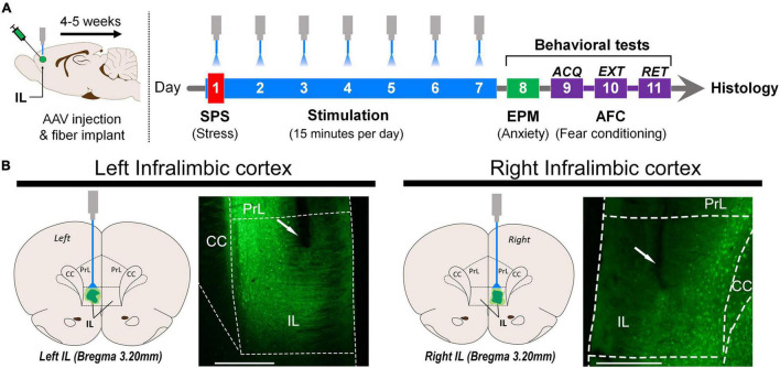
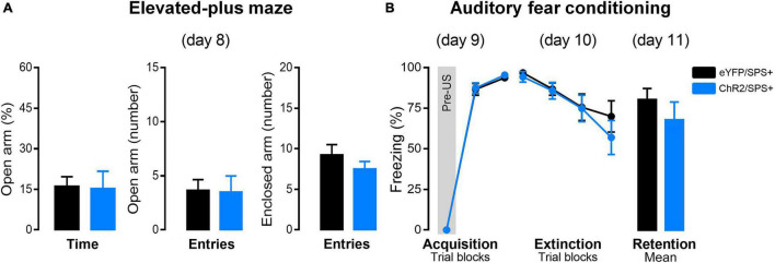
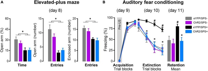
Post-traumatic stress disorder (PTSD) is associated with decreased activity in the prefrontal cortex. PTSD-like pathophysiology and behaviors have been observed in rodents exposed to a single prolonged stress (SPS) procedure. When animals are left alone for 7 days after SPS treatment, they show increased anxiety-like behavior and impaired extinction of conditioned fear, and reduced activity in the prefrontal cortex. Here, we tested the hypothesis that daily optogenetic stimulation of the infralimbic region (IL) of the medial prefrontal cortex (mPFC) during the 7 days after SPS would reverse SPS effects on anxiety and fear extinction. Male Sprague-Dawley rats underwent SPS and then received daily optogenetic stimulation (20 Hz, 2 s trains, every 10 s for 15 min/day) of glutamatergic neurons of the left or right IL for seven days. After this incubation period, rats were tested in the elevated plus-maze (EPM). Twenty-four hours after the EPM test, rats underwent auditory fear conditioning (AFC), extinction training and a retention test. SPS increased anxiety-like behavior in the EPM task and produced a profound impairment in extinction of AFC. Optogenetic stimulation of the left IL, but not right, during the 7-day incubation period reversed the extinction impairment. Optogenetic stimulation did not reverse the increased anxiety-like behavior, suggesting that the extinction effects are not due to a treatment-induced reduction in anxiety. Results indicate that increased activity of the left IL after traumatic experiences can prevent development of extinction impairments. These findings suggest that non-invasive brain stimulation may be a useful tool for preventing maladaptive responses to trauma.
创伤后应激障碍(PTSD)与前额叶皮质活动减少有关。在经历单次长时间应激(SPS)程序的啮齿动物中观察到了类似PTSD的病理生理学和行为表现。在SPS处理后让动物单独饲养7天,它们会表现出焦虑样行为增加、条件性恐惧消退受损以及前额叶皮质活动减少。在此,我们检验了这样一个假设:在SPS后的7天里,每天对内侧前额叶皮质(mPFC)的下缘区(IL)进行光遗传学刺激,将逆转SPS对焦虑和恐惧消退的影响。雄性Sprague-Dawley大鼠接受SPS处理,然后在7天时间里每天对左侧或右侧IL的谷氨酸能神经元进行光遗传学刺激(20赫兹,2秒脉冲串,每10秒一次,每次15分钟/天)。在这个潜伏期过后,大鼠在高架十字迷宫(EPM)中接受测试。在EPM测试24小时后,大鼠接受听觉恐惧条件反射(AFC)、消退训练和记忆测试。SPS增加了EPM任务中的焦虑样行为,并在AFC消退方面产生了严重损害。在7天潜伏期内对左侧IL而非右侧IL进行光遗传学刺激,逆转了消退损害。光遗传学刺激并未逆转增加的焦虑样行为,这表明消退效应并非由于治疗导致的焦虑减少。结果表明,创伤经历后左侧IL活动增加可以预防消退损害的发展。这些发现表明,非侵入性脑刺激可能是预防对创伤的适应不良反应的一种有用工具。