Urraro Fabrizio, Nardone Valerio, Reginelli Alfonso, Varelli Carlo, Angrisani Antonio, Patanè Vittorio, D'Ambrosio Luca, Roccatagliata Pietro, Russo Gaetano Maria, Gallo Luigi, De Chiara Marco, Altucci Lucia, Cappabianca Salvatore
Department of Precision Medicine, University of Campania Luigi Vanvitelli, Naples, Italy.
Istituto Diagnostico Varelli, Naples, Italy.
Front Oncol. 2021 Dec 21;11:805137. doi: 10.3389/fonc.2021.805137. eCollection 2021.
Radiomics can provide quantitative features from medical imaging that can be correlated to clinical endpoints. The challenges relevant to robustness of radiomics features have been analyzed by many researchers, as it seems to be influenced by acquisition and reconstruction protocols, as well as by the segmentation of the region of interest (ROI). Prostate cancer (PCa) represents a difficult playground for this technique, due to discrepancies in the identification of the cancer lesion and the heterogeneity of the acquisition protocols. The aim of this study was to investigate the reliability of radiomics in PCa magnetic resonance imaging (MRI).
A homogeneous cohort of patients with a PSA rise that underwent multiparametric MRI imaging of the prostate before biopsy was tested in this study. All the patients were acquired with the same MRI scanner, with a standardized protocol. The identification and the contouring of the region of interest (ROI) of an MRI suspicious cancer lesion were done by two radiologists with great experience in prostate cancer (>10 years). After the segmentation, the texture features were extracted with LIFEx. Texture features were then tested with intraclass coefficient correlation (ICC) analysis to analyze the reliability of the segmentation.
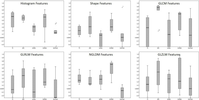
Forty-four consecutive patients were included in the present analysis. In 26 patients (59.1%), the prostate biopsy confirmed the presence of prostate cancer, which was scored as Gleason 6 in 6 patients (13.6%), Gleason 3 + 4 in 8 patients (18.2%), and Gleason 4 + 3 in 12 patients (27.3%). The reliability analysis conversely showed poor reliability in the majority of the MRI acquisition (61% in T2, 89% in DWI50, 44% in DWI400, and 83% in DWI1,500), with ADC acquisition only showing better reliability (poor reliability in only 33% of the texture features).
The low ratio of reliability in a monoinstitutional homogeneous cohort represents a significant alarm bell for the application of MRI radiomics in the field of prostate cancer. More work is needed in a clinical setting to further study the potential of MRI radiomics in prostate cancer.
放射组学可从医学影像中提供可与临床终点相关联的定量特征。许多研究人员已分析了与放射组学特征稳健性相关的挑战,因为其似乎受采集和重建协议以及感兴趣区域(ROI)分割的影响。由于癌症病灶识别存在差异以及采集协议的异质性,前列腺癌(PCa)是该技术的一个难题领域。本研究的目的是调查放射组学在PCa磁共振成像(MRI)中的可靠性。
本研究测试了一组在活检前接受前列腺多参数MRI成像且PSA升高的同质患者队列。所有患者均使用同一台MRI扫描仪,并采用标准化协议进行采集。由两位在前列腺癌方面有丰富经验(超过10年)的放射科医生对MRI可疑癌症病灶的感兴趣区域(ROI)进行识别和勾勒轮廓。分割后,使用LIFEx提取纹理特征。然后通过组内相关系数(ICC)分析对纹理特征进行测试,以分析分割的可靠性。
本分析纳入了44例连续患者。26例患者(59.1%)前列腺活检证实存在前列腺癌,其中6例患者(13.6%)评分为Gleason 6,8例患者(18.2%)评分为Gleason 3 + 4,12例患者(27.3%)评分为Gleason 4 + 3。相反,可靠性分析显示大多数MRI采集(T2加权像中为61%,DWI50序列中为89%,DWI400序列中为44%,DWI1500序列中为83%)的可靠性较差,只有ADC采集显示出较好的可靠性(仅33%的纹理特征可靠性较差)。
在单机构同质队列中可靠性比例较低,这对MRI放射组学在前列腺癌领域的应用来说是一个重大警示信号。在临床环境中需要开展更多工作,以进一步研究MRI放射组学在前列腺癌中的潜力。