• 文献检索
  • 文档翻译
  • 深度研究
  • 学术资讯
  • Suppr Zotero 插件Zotero 插件
  • 邀请有礼
  • 套餐&价格
  • 历史记录
应用&插件
Suppr Zotero 插件Zotero 插件浏览器插件Mac 客户端Windows 客户端微信小程序
定价
高级版会员购买积分包购买API积分包
服务
文献检索文档翻译深度研究API 文档MCP 服务
关于我们
关于 Suppr公司介绍联系我们用户协议隐私条款
关注我们

Suppr 超能文献

核心技术专利:CN118964589B侵权必究
粤ICP备2023148730 号-1Suppr @ 2026

文献检索

告别复杂PubMed语法,用中文像聊天一样搜索,搜遍4000万医学文献。AI智能推荐,让科研检索更轻松。

立即免费搜索

文件翻译

保留排版,准确专业,支持PDF/Word/PPT等文件格式,支持 12+语言互译。

免费翻译文档

深度研究

AI帮你快速写综述,25分钟生成高质量综述,智能提取关键信息,辅助科研写作。

立即免费体验

使用18 kDa转运蛋白PET根据先天免疫细胞激活对多发性硬化症病变进行表型分析。

Phenotyping of multiple sclerosis lesions according to innate immune cell activation using 18 kDa translocator protein-PET.

作者信息

Nylund Marjo, Sucksdorff Marcus, Matilainen Markus, Polvinen Eero, Tuisku Jouni, Airas Laura

机构信息

Turku PET Centre, Turku, Finland.

Clinical Neurosciences, University of Turku, Turku, Finland.

出版信息

Brain Commun. 2021 Dec 22;4(1):fcab301. doi: 10.1093/braincomms/fcab301. eCollection 2022 Feb.

DOI:10.1093/braincomms/fcab301
PMID:34993478
原文链接:https://pmc.ncbi.nlm.nih.gov/articles/PMC8727984/
Abstract

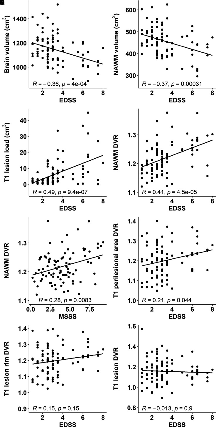
Chronic active lesions are promotors of neurodegeneration and disease progression in multiple sclerosis. They harbour a dense rim of activated innate immune cells at the lesion edge, which promotes lesion growth and thereby induces damage. Conventional MRI is of limited help in identifying the chronic active lesions, so alternative imaging modalities are needed. Objectives were to develop a PET-based automated analysis method for phenotyping of chronic lesions based on lesion-associated innate immune cell activation and to comprehensively evaluate the prevalence of these lesions in the various clinical subtypes of multiple sclerosis, and their association with disability. In this work, we use 18 kDa translocator protein-PET imaging for phenotyping chronic multiple sclerosis lesions at a large scale. For this, we identified 1510 white matter T1-hypointense lesions from 91 multiple sclerosis patients (67 relapsing-remitting patients and 24 secondary progressive patients). Innate immune cell activation at the lesion rim was measured using PET imaging and the 18 kDa translocator protein-binding radioligand C-PK11195. A T1-hypointense lesion was classified as rim-active if the distribution volume ratio of C-PK11195-binding was low in the plaque core and considerably higher at the plaque edge. If no significant ligand binding was observed, the lesion was classified as inactive. Plaques that had considerable ligand binding both in the core and at the rim were classified as overall-active. Conventional MRI and disability assessment using the Expanded Disability Status Scale were performed at the time of PET imaging. In the secondary progressive cohort, an average of 19% (median, interquartile range: 11-26) of T1 lesions were rim-active in each individual patient, compared to 10% (interquartile range: 0-20) among relapsing-remitting patients ( = 0.009). Secondary progressive patients had a median of 3 (range: 0-11) rim-active lesions, versus 1 (range: 0-18) among relapsing-remitting patients ( = 0.029). Among those patients who had rim-active lesions ( = 63), the average number of active voxels at the rim was higher among secondary progressive compared to relapsing-remitting patients (median 158 versus 74; = 0.022). The number of active voxels at the rim correlated significantly with the Expanded Disability Status Scale ( = 0.43, < 0.001), and the volume of the rim-active lesions similarly correlated with the Expanded Disability Status Scale ( = 0.45, < 0.001). Our study is the first to report phenotyping of chronic lesions at large scale, based on 18 kDa translocator protein-PET. Patients with higher disability displayed a higher proportion of rim-active lesions. The lesion phenotyping methodology offers a new tool for individual assessment of smouldering (rim-active) lesion burden.

摘要
https://cdn.ncbi.nlm.nih.gov/pmc/blobs/0bb8/8727984/ac9d3b99bdec/fcab301f6.jpg
https://cdn.ncbi.nlm.nih.gov/pmc/blobs/0bb8/8727984/c4a8f2a03da4/fcab301ga1.jpg
https://cdn.ncbi.nlm.nih.gov/pmc/blobs/0bb8/8727984/d02790688f07/fcab301f1.jpg
https://cdn.ncbi.nlm.nih.gov/pmc/blobs/0bb8/8727984/e37892fa4f92/fcab301f2.jpg
https://cdn.ncbi.nlm.nih.gov/pmc/blobs/0bb8/8727984/aa9a96dcdadf/fcab301f3.jpg
https://cdn.ncbi.nlm.nih.gov/pmc/blobs/0bb8/8727984/1df635348861/fcab301f4.jpg
https://cdn.ncbi.nlm.nih.gov/pmc/blobs/0bb8/8727984/ad2cda6c71ad/fcab301f5.jpg
https://cdn.ncbi.nlm.nih.gov/pmc/blobs/0bb8/8727984/ac9d3b99bdec/fcab301f6.jpg

慢性活动性病灶是多发性硬化症中神经退行性变和疾病进展的促进因素。它们在病灶边缘有一层密集的活化固有免疫细胞,这会促进病灶生长,从而导致损伤。传统的磁共振成像在识别慢性活动性病灶方面帮助有限,因此需要其他成像方式。目的是开发一种基于正电子发射断层扫描(PET)的自动分析方法,用于基于病灶相关固有免疫细胞活化对慢性病灶进行表型分析,并全面评估这些病灶在多发性硬化症各种临床亚型中的患病率及其与残疾的关联。在这项研究中,我们使用18 kDa转位蛋白PET成像对慢性多发性硬化症病灶进行大规模表型分析。为此,我们从91例多发性硬化症患者(67例复发缓解型患者和24例继发进展型患者)中识别出1510个白质T1低信号病灶。使用PET成像和18 kDa转位蛋白结合放射性配体C-PK11195测量病灶边缘的固有免疫细胞活化情况。如果C-PK11195结合的分布容积比在斑块核心较低而在斑块边缘显著较高,则T1低信号病灶被分类为边缘活化。如果未观察到明显的配体结合,则该病灶被分类为非活化。在核心和边缘均有相当数量配体结合的斑块被分类为整体活化。在PET成像时进行传统的磁共振成像以及使用扩展残疾状态量表进行残疾评估。在继发进展型队列中,每位患者平均有19%(中位数,四分位间距:11%-26%)的T1病灶为边缘活化,而复发缓解型患者中这一比例为10%(四分位间距:0%-20%)(P = 0.009)。继发进展型患者边缘活化病灶的中位数为3个(范围:0-11个),而复发缓解型患者为1个(范围:0-18个)(P = 0.029)。在那些有边缘活化病灶的患者中(n = 63),继发进展型患者边缘活化体素的平均数高于复发缓解型患者(中位数分别为158和74;P = 0.022)。边缘活化体素的数量与扩展残疾状态量表显著相关(P = 0.43,P < 0.001),边缘活化病灶的体积与扩展残疾状态量表同样显著相关(P = 0.45,P < 0.001)。我们的研究首次报告了基于18 kDa转位蛋白PET对慢性病灶进行大规模表型分析。残疾程度较高的患者边缘活化病灶的比例更高。这种病灶表型分析方法为个体评估隐匿性(边缘活化)病灶负担提供了一种新工具。

相似文献

1
Phenotyping of multiple sclerosis lesions according to innate immune cell activation using 18 kDa translocator protein-PET.使用18 kDa转运蛋白PET根据先天免疫细胞激活对多发性硬化症病变进行表型分析。
Brain Commun. 2021 Dec 22;4(1):fcab301. doi: 10.1093/braincomms/fcab301. eCollection 2022 Feb.
2
Contribution of new and chronic cortical lesions to disability accrual in multiple sclerosis.新的和慢性皮质病变对多发性硬化症残疾累积的影响。
Brain Commun. 2024 May 2;6(3):fcae158. doi: 10.1093/braincomms/fcae158. eCollection 2024.
3
Longitudinal stability of progression-related microglial activity during teriflunomide treatment in patients with multiple sclerosis.多发性硬化症患者用特立氟胺治疗期间与疾病进展相关的小胶质细胞活性的纵向稳定性。
Eur J Neurol. 2023 Aug;30(8):2365-2375. doi: 10.1111/ene.15834. Epub 2023 May 23.
4
Quantitative susceptibility mapping identifies inflammation in a subset of chronic multiple sclerosis lesions.定量磁化率映射可识别慢性多发性硬化症病变亚群中的炎症。
Brain. 2019 Jan 1;142(1):133-145. doi: 10.1093/brain/awy296.
5
Positron Emission Tomography with [ F]-DPA-714 Unveils a Smoldering Component in Most Multiple Sclerosis Lesions which Drives Disease Progression.正电子发射断层扫描(PET)结合 [ F]-DPA-714 揭示了大多数多发性硬化病变中的潜伏成分,该成分驱动疾病进展。
Ann Neurol. 2023 Aug;94(2):366-383. doi: 10.1002/ana.26657. Epub 2023 May 3.
6
Long-term evolution of multiple sclerosis iron rim lesions in 7 T MRI.7T MRI 下多发性硬化症铁环病变的长期演变。
Brain. 2021 Apr 12;144(3):833-847. doi: 10.1093/brain/awaa436.
7
Association of serum neurofilament light with microglial activation in multiple sclerosis.血清神经丝轻链与多发性硬化症中微胶质细胞激活的关联。
J Neurol Neurosurg Psychiatry. 2023 Sep;94(9):698-706. doi: 10.1136/jnnp-2023-331051. Epub 2023 May 2.
8
Cortical and phase rim lesions on 7 T MRI as markers of multiple sclerosis disease progression.7T磁共振成像上的皮质和相缘病变作为多发性硬化疾病进展的标志物
Brain Commun. 2021 Jun 24;3(3):fcab134. doi: 10.1093/braincomms/fcab134. eCollection 2021.
9
Neuroinflammation and its relationship to changes in brain volume and white matter lesions in multiple sclerosis.神经炎症及其与多发性硬化症脑容量变化和白质病变的关系。
Brain. 2017 Nov 1;140(11):2927-2938. doi: 10.1093/brain/awx228.
10
Chronic white matter lesion activity predicts clinical progression in primary progressive multiple sclerosis.慢性白质病变活动可预测原发性进行性多发性硬化症的临床进展。
Brain. 2019 Sep 1;142(9):2787-2799. doi: 10.1093/brain/awz212.

引用本文的文献

1
Longitudinal accumulation of glial activation measured by TSPO-PET predicts later brain atrophy in multiple sclerosis.通过TSPO-PET测量的胶质细胞激活的纵向积累可预测多发性硬化症患者后期的脑萎缩。
J Neuroinflammation. 2025 Aug 7;22(1):200. doi: 10.1186/s12974-025-03519-y.
2
Serum glial fibrillary acid protein associates with TSPO-expressing lesions in multiple sclerosis brain.血清胶质纤维酸性蛋白与多发性硬化症脑部表达TSPO的病灶相关。
Ther Adv Neurol Disord. 2025 Jul 28;18:17562864251352998. doi: 10.1177/17562864251352998. eCollection 2025.
3
Innate Immune Mechanisms in the Central Nervous System.

本文引用的文献

1
Long-term evolution of multiple sclerosis iron rim lesions in 7 T MRI.7T MRI 下多发性硬化症铁环病变的长期演变。
Brain. 2021 Apr 12;144(3):833-847. doi: 10.1093/brain/awaa436.
2
Cellular sources of TSPO expression in healthy and diseased brain.健康和患病大脑中 TSPO 表达的细胞来源。
Eur J Nucl Med Mol Imaging. 2021 Dec;49(1):146-163. doi: 10.1007/s00259-020-05166-2. Epub 2021 Jan 12.
3
Paramagnetic Rims in Multiple Sclerosis and Neuromyelitis Optica Spectrum Disorder: A Quantitative Susceptibility Mapping Study with 3-T MRI.
中枢神经系统中的固有免疫机制
Adv Exp Med Biol. 2025;1476:381-409. doi: 10.1007/978-3-031-85340-1_15.
4
Determinants driving the evolution of new multiple sclerosis lesions into chronic active or remyelinated states.促使新的多发性硬化症病灶演变为慢性活动状态或再髓鞘化状态的决定因素。
Neuroimage Clin. 2025 Jun 10;47:103823. doi: 10.1016/j.nicl.2025.103823.
5
The role of microglia in multiple sclerosis: implications for treatment with Bruton's tyrosine kinase inhibitors.小胶质细胞在多发性硬化症中的作用:对布鲁顿酪氨酸激酶抑制剂治疗的启示。
Front Immunol. 2025 May 15;16:1495529. doi: 10.3389/fimmu.2025.1495529. eCollection 2025.
6
Biomarkers of Progression Independent of Relapse Activity-Can We Actually Measure It Yet?独立于复发活动的疾病进展生物标志物——我们真的能够对其进行测量了吗?
Int J Mol Sci. 2025 May 14;26(10):4704. doi: 10.3390/ijms26104704.
7
Associations between chronic active lesions and clinical outcomes in multiple sclerosis: A systematic literature review.多发性硬化症中慢性活动性病变与临床结局的关联:一项系统文献综述。
J Manag Care Spec Pharm. 2025 Jul;31(7):694-721. doi: 10.18553/jmcp.2025.24294. Epub 2025 May 13.
8
Broad rim lesions are a new pathological and imaging biomarker for rapid disease progression in multiple sclerosis.宽边病变是多发性硬化症快速疾病进展的一种新的病理和影像学生物标志物。
Nat Med. 2025 Apr 29. doi: 10.1038/s41591-025-03625-7.
9
Integrating TSPO-PET imaging with metabolomics for enhanced prognostic accuracy in multiple sclerosis.整合TSPO-PET成像与代谢组学以提高多发性硬化症的预后准确性。
BMJ Neurol Open. 2025 Apr 16;7(1):e001026. doi: 10.1136/bmjno-2025-001026. eCollection 2025.
10
TSPO-PET Reveals Higher Inflammation in White Matter Disrupted by Paramagnetic Rim Lesions in Multiple Sclerosis.TSPO-PET显示多发性硬化症中被顺磁边缘病变破坏的白质炎症更高。
bioRxiv. 2025 Jan 3:2025.01.03.627857. doi: 10.1101/2025.01.03.627857.
多发性硬化症和视神经脊髓炎谱系障碍中的顺磁性边缘:一项使用3-T MRI的定量磁化率映射研究
J Clin Neurol. 2020 Oct;16(4):562-572. doi: 10.3988/jcn.2020.16.4.562.
4
Brain TSPO-PET predicts later disease progression independent of relapses in multiple sclerosis.脑 TSPO-PET 预测多发性硬化症的疾病进展与复发无关。
Brain. 2020 Dec 5;143(11):3318-3330. doi: 10.1093/brain/awaa275.
5
Sex-specific differences in rim appearance of multiple sclerosis lesions on quantitative susceptibility mapping.基于定量磁化率图谱的多发性硬化症病灶边缘外观的性别差异
Mult Scler Relat Disord. 2020 Oct;45:102317. doi: 10.1016/j.msard.2020.102317. Epub 2020 Jun 18.
6
Evaluation of the 'ring sign' and the 'core sign' as a magnetic resonance imaging marker of disease activity and progression in clinically isolated syndrome and early multiple sclerosis.评估“环征”和“核心征”作为临床孤立综合征和早期多发性硬化症疾病活动及进展的磁共振成像标志物。
Mult Scler J Exp Transl Clin. 2020 Mar 30;6(1):2055217320915480. doi: 10.1177/2055217320915480. eCollection 2020 Jan-Mar.
7
Insights into disseminated MS brain pathology with multimodal diffusion tensor and PET imaging.多模态弥散张量和 PET 成像对多发性硬化脑病理学的研究进展。
Neurol Neuroimmunol Neuroinflamm. 2020 Mar 2;7(3). doi: 10.1212/NXI.0000000000000691. Print 2020 May.
8
Folate receptor-targeted positron emission tomography of experimental autoimmune encephalomyelitis in rats.叶酸受体靶向正电子发射断层扫描在大鼠实验性自身免疫性脑脊髓炎中的应用。
J Neuroinflammation. 2019 Dec 3;16(1):252. doi: 10.1186/s12974-019-1612-3.
9
The P2X receptor tracer [C]SMW139 as an in vivo marker of neuroinflammation in multiple sclerosis: a first-in man study.P2X受体示踪剂[C]SMW139作为多发性硬化症神经炎症的体内标志物:一项人体首次研究。
Eur J Nucl Med Mol Imaging. 2020 Feb;47(2):379-389. doi: 10.1007/s00259-019-04550-x. Epub 2019 Nov 8.
10
A quantitative neuropathological assessment of translocator protein expression in multiple sclerosis.多发性硬化症中转位蛋白表达的定量神经病理学评估。
Brain. 2019 Nov 1;142(11):3440-3455. doi: 10.1093/brain/awz287.