Blindenbacher Nelly, Brunner Eveline, Asseyer Susanna, Scheel Michael, Siebert Nadja, Rasche Ludwig, Bellmann-Strobl Judith, Brandt Alexander, Ruprecht Klemens, Meier Dominik, Wuerfel Jens, Paul Friedemann, Sinnecker Tim
qbig, Department of Biomedical Engineering, University of Basel, Switzerland.
Neurocure Clinical Research Center, Charité-Universitätsmedizin Berlin, Germany.
Mult Scler J Exp Transl Clin. 2020 Mar 30;6(1):2055217320915480. doi: 10.1177/2055217320915480. eCollection 2020 Jan-Mar.
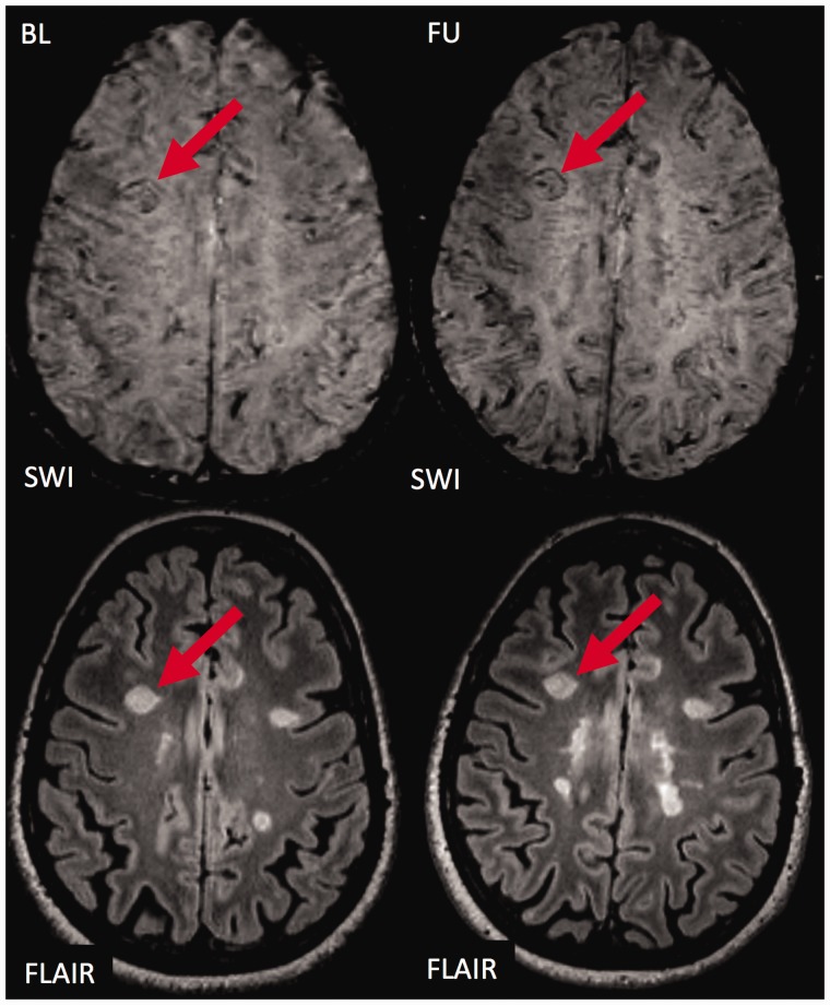
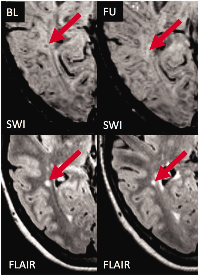
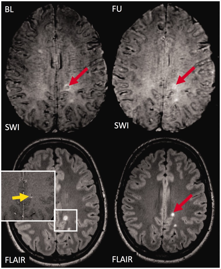
Brain lesions with a hypointense ring or core were described in multiple sclerosis on susceptibility weighted imaging.
The purpose of this study was to study the evolution and prognostic relevance of susceptibility weighted imaging hypointense lesions in clinically isolated syndrome and early multiple sclerosis.
Sixty-six early multiple sclerosis and clinically isolated syndrome patients were followed over a median period of 2.9 years (range 1.6-4.6 years) and underwent 3T magnetic resonance imaging including 3D susceptibility weighted imaging and T2-weighted fluid-attenuated inversion recovery. We assessed the presence of susceptibility weighted imaging hypointense core or ring lesions, and Expanded Disability Status Scale at baseline and follow-up.
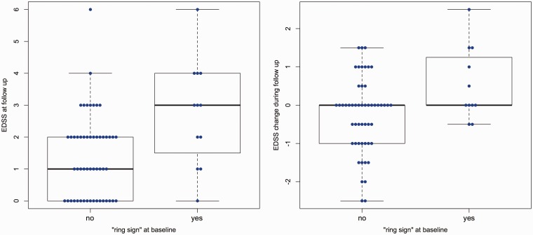
Of 611 lesions at baseline, 64 (10.5%) had a susceptibility weighted imaging hypointense core, and 28 (4.6%) had a susceptibility weighted imaging hypointense ring. Hypointense ring lesions were larger ( < 0.001) and more T1w hypointense ( = 0.002) than others. During follow-up, hypointense core lesions became susceptibility weighted imaging isointense (52 lesions, 81%); few developed into hypointense ring lesions (two lesions, 3%). Hypointense ring lesions did not shrink on T2-weighted fluid-attenuated inversion recovery images ( = 0.077, trend towards more enlargement compared to others), while hypointense core lesions more often shrunk in comparison to lesions without a hypointense core ( = 0.002). The number of susceptibility weighted imaging hypointense ring lesions at baseline correlated with Expanded Disability Status Scale progression at follow-up ( = 0.021, R = 0.289).
In our cohort of patients with clinically isolated syndrome or early multiple sclerosis, susceptibility weighted imaging hypointense ring lesions were only rarely detectable, but did not shrink and were associated with future disability progression.
在多系统硬化症的磁敏感加权成像中发现了具有低信号环或核心的脑病变。
本研究旨在探讨临床孤立综合征和早期多系统硬化症中磁敏感加权成像低信号病变的演变及其与预后的相关性。
对66例早期多系统硬化症和临床孤立综合征患者进行了为期2.9年(范围1.6 - 4.6年)的随访,并接受了3T磁共振成像检查,包括三维磁敏感加权成像和T2加权液体衰减反转恢复序列成像。我们评估了磁敏感加权成像低信号核心或环病变的存在情况,以及基线和随访时的扩展残疾状态量表。
在基线时的611个病变中,64个(10.5%)有磁敏感加权成像低信号核心,28个(4.6%)有磁敏感加权成像低信号环。低信号环病变比其他病变更大(<0.001)且T1加权像上低信号更明显(=0.002)。在随访期间,低信号核心病变在磁敏感加权成像上变为等信号(52个病变,81%);少数发展为低信号环病变(2个病变,3%)。低信号环病变在T2加权液体衰减反转恢复序列图像上没有缩小(=0.077,与其他病变相比有增大趋势),而低信号核心病变与无低信号核心的病变相比更常缩小(=0.002)。基线时磁敏感加权成像低信号环病变的数量与随访时扩展残疾状态量表的进展相关(=0.021,R = 0.