Lagan Jakub, Naish Josephine H, Bradley Joshua, Fortune Christien, Palmer Charlie, Clark David, Schelbert Erik B, Schmitt Matthias, Bright-Thomas Rowland, Miller Christopher A
Manchester University NHS Foundation Trust, Wythenshawe Hospital, Southmoor Road, Wythenshawe, Manchester, M23 9LT, England, UK.
Division of Cardiovascular Sciences, School of Medical Sciences, Faculty of Biology, Medicine and Health, Manchester Academic Health Science Centre, University of Manchester, Oxford Road, Manchester, M13 9PL, England, UK.
Int J Cardiovasc Imaging. 2022 May;38(5):1121-1131. doi: 10.1007/s10554-021-02496-6. Epub 2022 Jan 7.
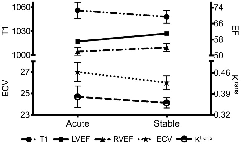
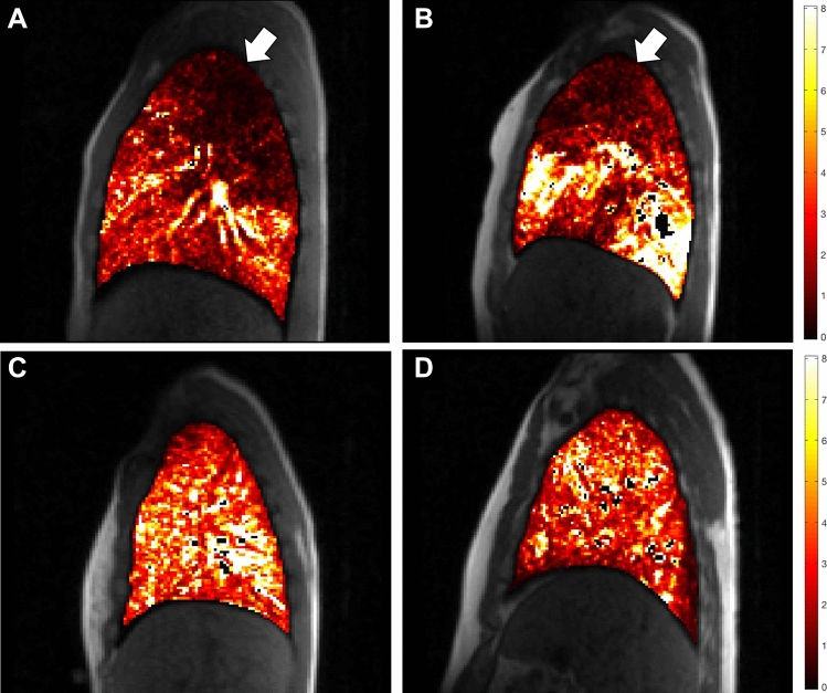
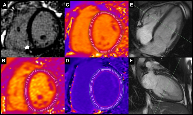
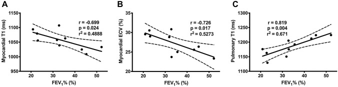
Cystic fibrosis (CF) transmembrane conductance regulator is expressed in myocardium, but cardiac involvement in CF remains poorly understood. The recent development of a combined cardiopulmonary magnetic resonance imaging technology allows for a simultaneous interrogation of cardiac and pulmonary structure and function. The aim of this study was to investigate myocardial manifestations in adults with CF, both in a stable state and during an acute respiratory exacerbation, and to investigate the relationship between cardiac and pulmonary disease. Healthy adult volunteers (n = 12) and adults with CF (n = 10) were studied using a multiparametric cardiopulmonary magnetic resonance protocol. CF patients were scanned during an acute respiratory exacerbation and re-scanned when stable. Stable CF was associated with left ventricular dilatation and hypertrophy (LVH; left ventricular mass: CF 59 ± 9 g/m vs. control 50 ± 8 g/m; p = 0.028). LVH was predominantly driven by extracellular myocardial matrix expansion (extracellular matrix mass: CF 27.5 ± 3.4 g vs. control 23.6 ± 5.2 g; p = 0.006; extracellular volume [ECV]: CF 27.6 [24.7-29.8]% vs. control 24.8 [22.9-26.0]%; p = 0.030). Acute CF was associated with an acute reduction in left ventricular function (ejection fraction: acute 57 ± 3% vs. stable 61 ± 5%; p = 0.025) and there was a suggestion of myocardial oedema. Myocardial oedema severity was strongly associated with the severity of airflow limitation (r = - 0.726, p = 0.017). Multiparametric cardiopulmonary magnetic resonance technology allows for a simultaneous interrogation of cardiac and pulmonary structure and function. Stable CF is associated with adverse myocardial remodelling, including left ventricular systolic dilatation and hypertrophy, driven by myocardial fibrosis. CF exacerbation is associated with acute myocardial contractile dysfunction. There is also a suggestion of myocardial oedema in the acute period which is related to pulmonary disease severity.
囊性纤维化(CF)跨膜传导调节因子在心肌中表达,但CF患者的心脏受累情况仍知之甚少。最近开发的心肺联合磁共振成像技术能够同时检测心脏和肺部的结构与功能。本研究的目的是调查CF成年患者在稳定状态和急性呼吸加重期的心肌表现,并研究心脏与肺部疾病之间的关系。使用多参数心肺磁共振成像方案对健康成年志愿者(n = 12)和CF成年患者(n = 10)进行了研究。CF患者在急性呼吸加重期进行扫描,并在病情稳定时再次扫描。稳定期CF与左心室扩张和肥厚(LVH;左心室质量:CF组59±9 g/m vs.对照组50±8 g/m;p = 0.028)相关。LVH主要由细胞外心肌基质扩张驱动(细胞外基质质量:CF组27.5±3.4 g vs.对照组23.6±5.2 g;p = 0.006;细胞外容积[ECV]:CF组27.6[24.7 - 29.8]% vs.对照组24.8[22.9 - 26.0]%;p = 0.030)。急性CF与左心室功能急性下降相关(射血分数:急性发作期57±3% vs.稳定期