Department of Histology and Embryology, Poznan University of Life Sciences, Wojska Polskiego 71C, PL 60-625, Poznań, Poland.
BMC Vet Res. 2022 Jan 7;18(1):21. doi: 10.1186/s12917-021-03111-5.
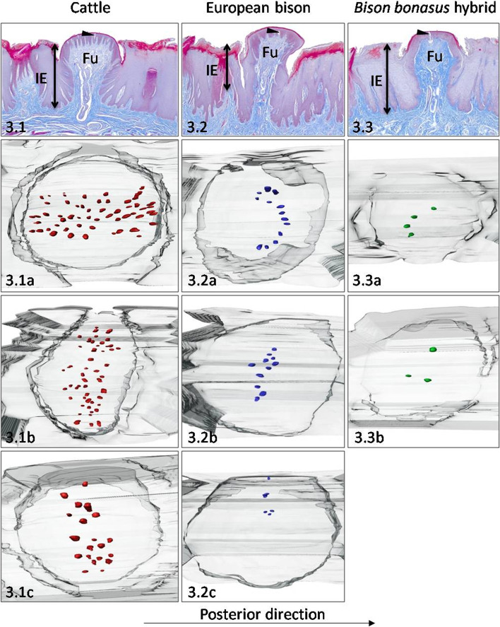
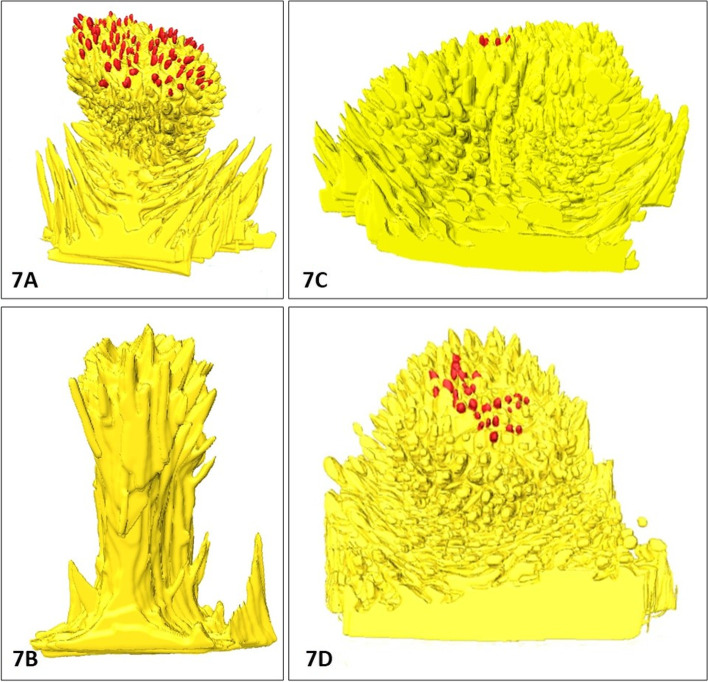
Our recent macro- and scanning electron microscopic study of tongue conducted on domesticated cattle, wild living European bison, and Bison bonasus hybrid revealed various spatial arrangement and number of gustatory and mechanical papillae between parental species and their hybrid. Furthermore, scanning electron microscopy analysis of gustatory papillae indicated the variable distribution of fungiform papillae (Fu) over the surface of the tongue, which could be significant in differentiated taste perception during feeding in studied wild living and domesticated husbandry ruminants. To specify the detailed microstructure of Fu papillae with connective tissue cores (CTC) and intraepithelial taste buds system, the first time the three-dimensional computer-aided analysis of serial histoslides resulted in the rendering of 3D reconstructions of Fu papillae.
The comparative analysis of 3D models Fu papillae conducted in six areas of lingual mucosa of each tongue revealed information about, microstructural diversity of Fu papillae in studied ruminants. The estimation of number and density of Fu papillae on tongues, rate of protrusion of papillae over mucosa, and a number of taste buds per papilla allowed to state the ventral surface of the lingual apex and posterolateral surfaces of the lingual torus as regions important in taste perception, as in the preselection of taken food, as well in the analysis of food during rumination, respectively. On the 3D models were observed three structural types of CTC of different distribution on the tongue in studied species. The quantitative data of the number of taste buds on Fu papillae have regional functional differences in the taste system important in feeding and veterinary practice. Moreover, our analysis determined specific features in examined hybrid and showed similarities of some studied features with cattle, i.e., maternal species.
The 3D reconstruction method used for the first time in the field of study of the lingual papillae and taste buds system can be considered as an innovative and effective tool in assessing of the microstructures of Fu papillae, and it could be suitable for further studies of taste system structures in normal and pathological condition.
我们最近对家养牛、野生欧洲野牛和野牛 bonasus 杂种的舌头进行了宏观和扫描电子显微镜研究,揭示了亲代物种及其杂种之间各种味觉和机械乳头的空间排列和数量。此外,对味觉乳头的扫描电子显微镜分析表明,在研究的野生和家养反刍动物的进食过程中,丝状乳头(Fu)在舌头上的分布存在可变性,这可能对味觉感知有重要意义。为了详细研究具有结缔组织核心(CTC)和上皮内味觉芽系统的 Fu 乳头的微观结构,我们首次对连续组织切片进行了三维计算机辅助分析,从而生成了 Fu 乳头的 3D 重建。
对每个舌头的舌黏膜六个区域的 Fu 乳头 3D 模型进行比较分析,揭示了研究反刍动物 Fu 乳头微观结构多样性的信息。对舌头上 Fu 乳头的数量和密度、乳头在黏膜上的突出率以及每个乳头的味蕾数进行估计,表明舌舌尖腹侧表面、舌嵴后外侧表面是味觉感知的重要区域,如在选择食物前,以及反刍过程中对食物的分析。在 3D 模型上观察到,在研究物种中,CTC 有三种不同分布的结构类型。在味觉系统中,Fu 乳头上的味蕾数量的定量数据在进食和兽医实践中具有重要的区域功能差异。此外,我们的分析确定了杂种中特定的特征,并表明了某些研究特征与母本物种牛的相似性。
首次在舌乳头和味觉芽系统研究领域中使用的 3D 重建方法可被视为评估 Fu 乳头微观结构的创新性和有效工具,并且可能适用于正常和病理条件下味觉系统结构的进一步研究。