Hinton and Garza Lopez Family Consulting Company, Iowa City, IA 52246, USA.
Department of Molecular Physiology and Biophysics, Vanderbilt University, Nashville, TN 37232, USA.
Cells. 2021 Dec 27;11(1):65. doi: 10.3390/cells11010065.
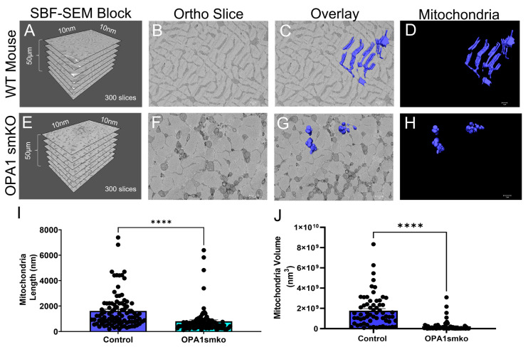
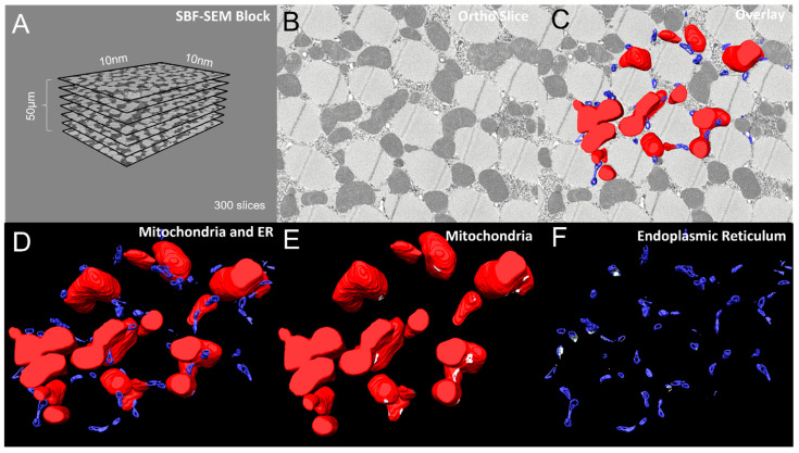
High-resolution 3D images of organelles are of paramount importance in cellular biology. Although light microscopy and transmission electron microscopy (TEM) have provided the standard for imaging cellular structures, they cannot provide 3D images. However, recent technological advances such as serial block-face scanning electron microscopy (SBF-SEM) and focused ion beam scanning electron microscopy (FIB-SEM) provide the tools to create 3D images for the ultrastructural analysis of organelles. Here, we describe a standardized protocol using the visualization software, Amira, to quantify organelle morphologies in 3D, thereby providing accurate and reproducible measurements of these cellular substructures. We demonstrate applications of SBF-SEM and Amira to quantify mitochondria and endoplasmic reticulum (ER) structures.
细胞器的高分辨率 3D 图像在细胞生物学中至关重要。尽管光学显微镜和透射电子显微镜(TEM)为细胞结构的成像提供了标准,但它们无法提供 3D 图像。然而,最近的技术进步,如连续块面扫描电子显微镜(SBF-SEM)和聚焦离子束扫描电子显微镜(FIB-SEM),为创建细胞器的 3D 图像提供了工具,用于超微结构分析。在这里,我们描述了一个使用可视化软件 Amira 的标准化方案,以定量 3D 中的细胞器形态,从而对这些细胞亚结构进行准确和可重复的测量。我们展示了 SBF-SEM 和 Amira 在定量线粒体和内质网(ER)结构方面的应用。