Department of Internal Medicine, Carver College of Medicine, University of Iowa, 375 Newton Rd, Iowa City, IA 52242, USA.
Fraternal Order of Eagles Diabetes Research Center, University of Iowa, 375 Newton Rd, Iowa City, IA 52242, USA.
Cells. 2021 Aug 24;10(9):2177. doi: 10.3390/cells10092177.
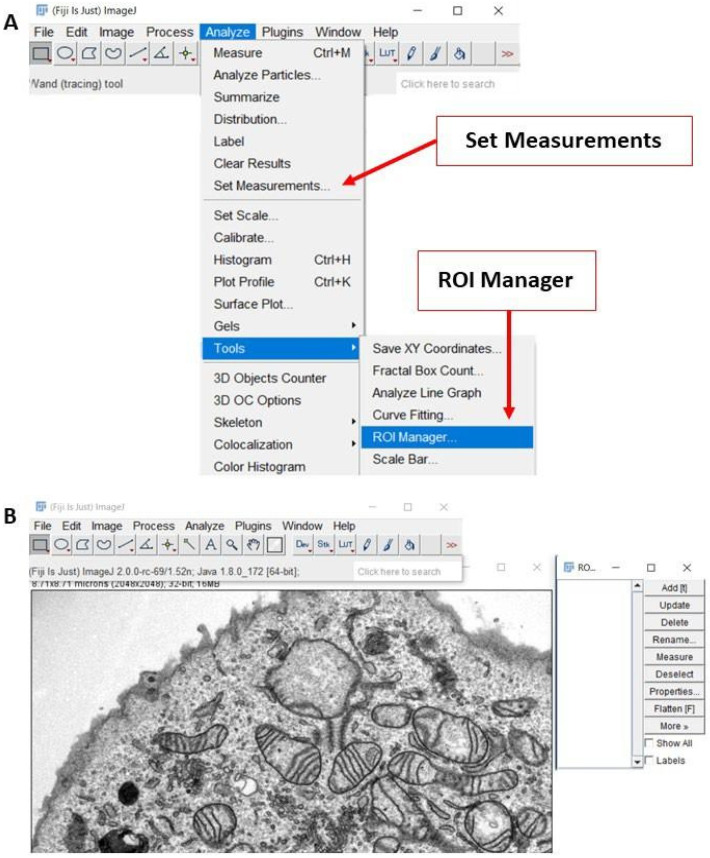
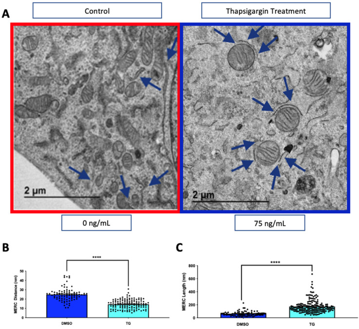
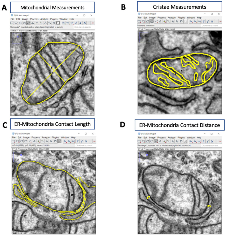
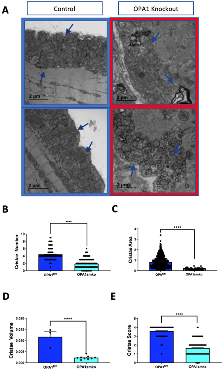
Transmission electron microscopy (TEM) is widely used as an imaging modality to provide high-resolution details of subcellular components within cells and tissues. Mitochondria and endoplasmic reticulum (ER) are organelles of particular interest to those investigating metabolic disorders. A straightforward method for quantifying and characterizing particular aspects of these organelles would be a useful tool. In this protocol, we outline how to accurately assess the morphology of these important subcellular structures using open source software , originally developed by the National Institutes of Health (NIH). Specifically, we detail how to obtain mitochondrial length, width, area, and circularity, in addition to assessing cristae morphology and measuring mito/endoplasmic reticulum (ER) interactions. These procedures provide useful tools for quantifying and characterizing key features of sub-cellular morphology, leading to accurate and reproducible measurements and visualizations of mitochondria and ER.
透射电子显微镜(TEM)广泛用作成像方式,以提供细胞和组织内亚细胞成分的高分辨率细节。线粒体和内质网(ER)是研究代谢紊乱的人特别感兴趣的细胞器。一种用于定量和表征这些细胞器特定方面的直接方法将是一种有用的工具。在本方案中,我们概述了如何使用开源软件准确评估这些重要亚细胞结构的形态,该软件最初由美国国立卫生研究院(NIH)开发。具体来说,我们详细说明了如何获得线粒体的长度、宽度、面积和圆形度,以及如何评估嵴形态和测量线粒体/内质网(ER)相互作用。这些程序为定量和表征亚细胞形态的关键特征提供了有用的工具,从而实现了对线粒体和 ER 的准确和可重复的测量和可视化。