Ruivo Carolina F, Bastos Nuno, Adem Barbara, Batista Ines, Duraes Cecilia, Melo Carlos A, Castaldo Stephanie A, Campos-Laborie Francisco, Moutinho-Ribeiro Pedro, Morão Barbara, Costa-Pinto Ana, Silva Soraia, Osorio Hugo, Ciordia Sergio, Costa Jose Luis, Goodrich David, Cavadas Bruno, Pereira Luisa, Kouzarides Tony, Macedo Guilherme, Maio Rui, Carneiro Fatima, Cravo Marília, Kalluri Raghu, Machado Jose Carlos, Melo Sonia A
i3S Instituto de Investigação e Inovação em Saúde, University of Porto, Porto, Portugal.
IPATIMUP Institute of Molecular Pathology and Immunology, University of Porto, Porto, Portugal.
Gut. 2022 Jan 10;71(10):2043-68. doi: 10.1136/gutjnl-2021-324994.
Intratumor heterogeneity drives cancer progression and therapy resistance. However, it has yet to be determined whether and how subpopulations of cancer cells interact and how this interaction affects the tumour.
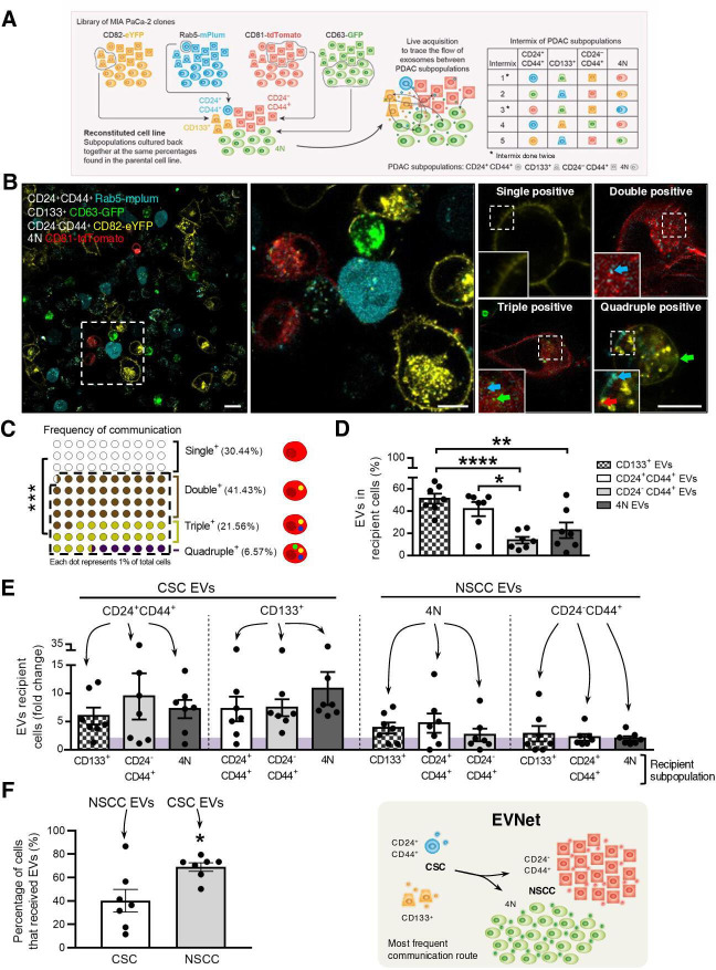
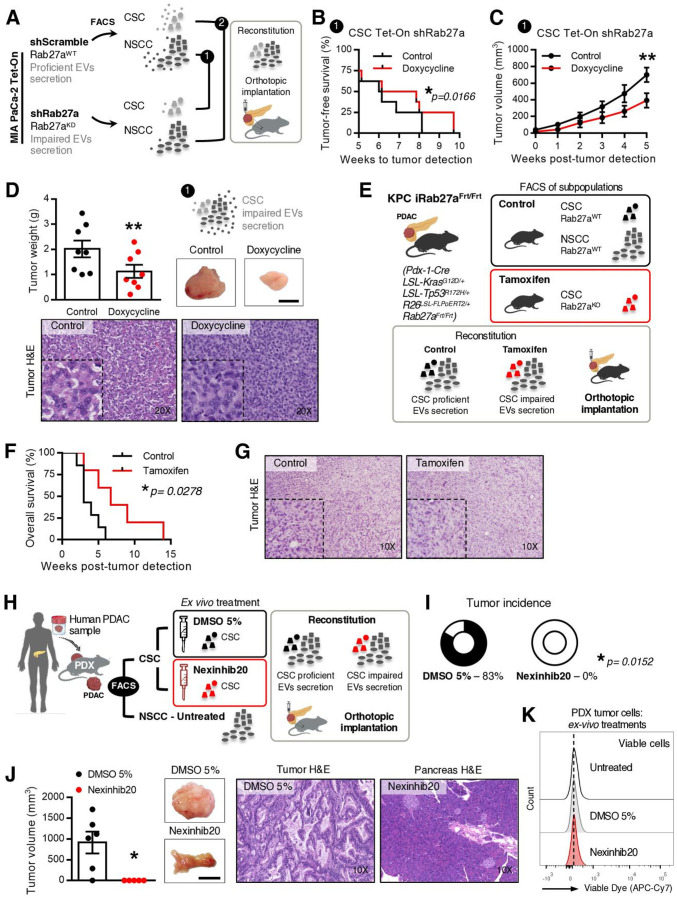
We have studied the spontaneous flow of extracellular vesicles (EVs) between subpopulations of cancer cells: cancer stem cells (CSC) and non-stem cancer cells (NSCC). To determine the biological significance of the most frequent communication route, we used pancreatic ductal adenocarcinoma (PDAC) orthotopic models, patient-derived xenografts (PDXs) and genetically engineered mouse models (GEMMs).
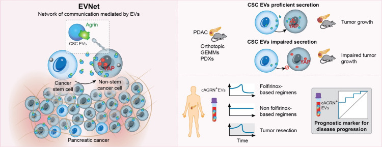
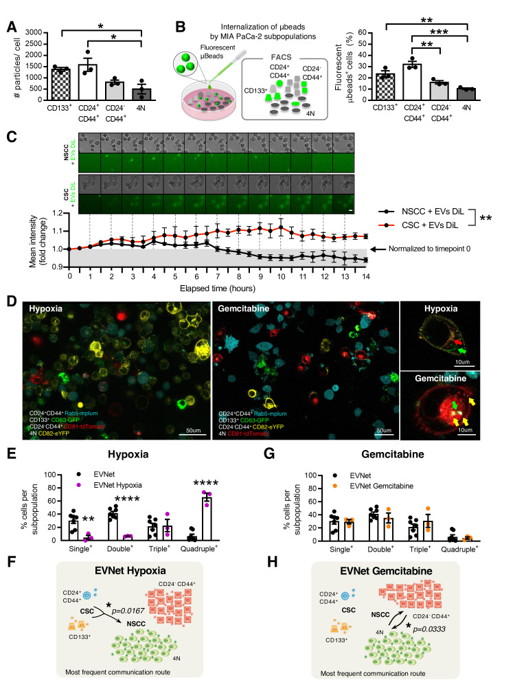
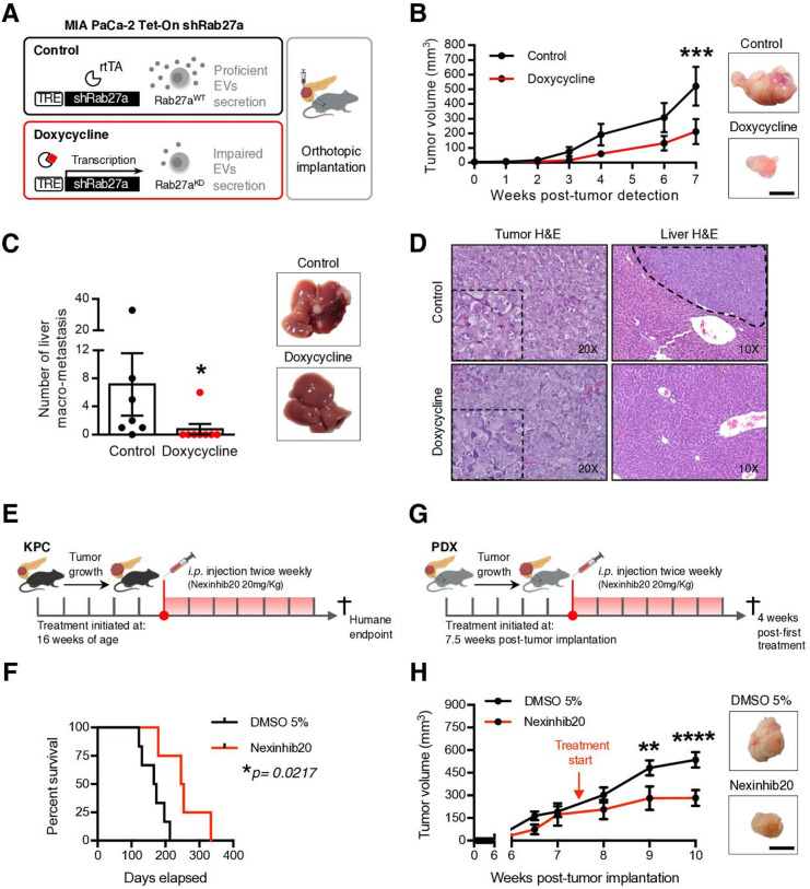
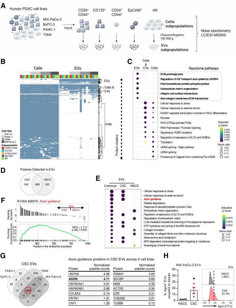
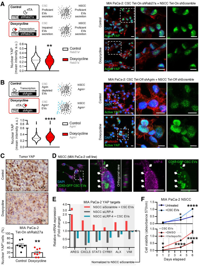
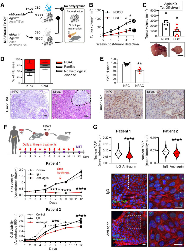
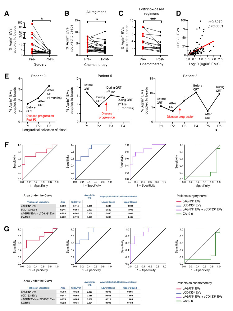
We demonstrate that PDAC tumours establish an organised communication network between subpopulations of cancer cells using EVs called the EVNet). The EVNet is plastic and reshapes in response to its environment. Communication within the EVNet occurs preferentially from CSC to NSCC. Inhibition of this communication route by impairing Rab27a function in orthotopic xenographs, GEMMs and PDXs is sufficient to hamper tumour growth and phenocopies the inhibition of communication in the whole tumour. Mechanistically, we provide evidence that CSC EVs use agrin protein to promote Yes1 associated transcriptional regulator (YAP) activation via LDL receptor related protein 4 (LRP-4). Ex vivo treatment of PDXs with antiagrin significantly impairs proliferation and decreases the levels of activated YAP.Patients with high levels of agrin and low inactive YAP show worse disease-free survival. In addition, patients with a higher number of circulating agrin EVs show a significant increased risk of disease progression.
PDAC tumours establish a cooperation network mediated by EVs that is led by CSC and agrin, which allows tumours to adapt and thrive. Targeting agrin could make targeted therapy possible for patients with PDAC and has a significant impact on CSC that feeds the tumour and is at the centre of therapy resistance.
肿瘤内异质性驱动癌症进展和治疗耐药性。然而,癌细胞亚群是否以及如何相互作用,以及这种相互作用如何影响肿瘤,目前尚待确定。
我们研究了癌细胞亚群(癌症干细胞[CSC]和非干细胞癌细胞[NSCC])之间细胞外囊泡(EV)的自发流动。为了确定最常见通讯途径的生物学意义,我们使用了胰腺导管腺癌(PDAC)原位模型、患者来源的异种移植模型(PDX)和基因工程小鼠模型(GEMM)。
我们证明,PDAC肿瘤利用称为EVNet的细胞外囊泡在癌细胞亚群之间建立了一个有组织的通讯网络。EVNet具有可塑性,并会根据其环境重塑。EVNet内的通讯优先从CSC到NSCC。通过损害原位异种移植、GEMM和PDX中的Rab27a功能来抑制这种通讯途径,足以阻碍肿瘤生长,并模拟对整个肿瘤通讯的抑制。从机制上讲,我们提供的证据表明,CSC来源的细胞外囊泡利用聚集蛋白通过低密度脂蛋白受体相关蛋白4(LRP-4)促进Yes1相关转录调节因子(YAP)的激活。用抗聚集蛋白对PDX进行体外处理可显著损害增殖并降低活化YAP的水平。聚集蛋白水平高且YAP非活性低的患者无病生存期较差。此外,循环中聚集蛋白来源的细胞外囊泡数量较多的患者疾病进展风险显著增加。
PDAC肿瘤建立了一个由CSC和聚集蛋白介导的细胞外囊泡合作网络,使肿瘤能够适应并茁壮成长。靶向聚集蛋白可能使PDAC患者的靶向治疗成为可能,并对滋养肿瘤且处于治疗耐药核心的CSC产生重大影响。