Cao Meng-Shu, Zhao Ting-Yan, Song Zhi-Long, Lu Hong-Ting, Zheng Yun, Gu Xiao-Ming, Lu Tao, Wang Qiong, Zhou Jing-Jun
Department of Cardiology, Xijing hospital, Fourth Military Medical University, Xi'an, 710032, China.
Department of Physiology and Pathophysiology, Fourth Military Medical University, Xi'an, 710032, China.
Cell Death Discov. 2022 Jan 10;8(1):10. doi: 10.1038/s41420-021-00810-8.
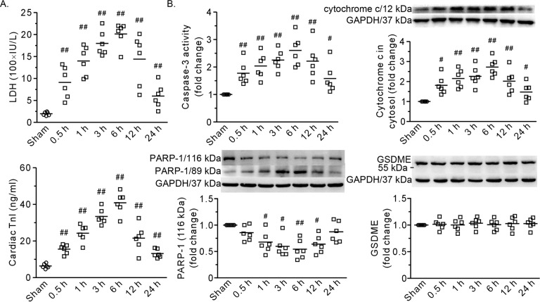
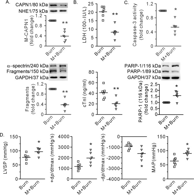
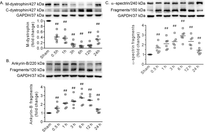
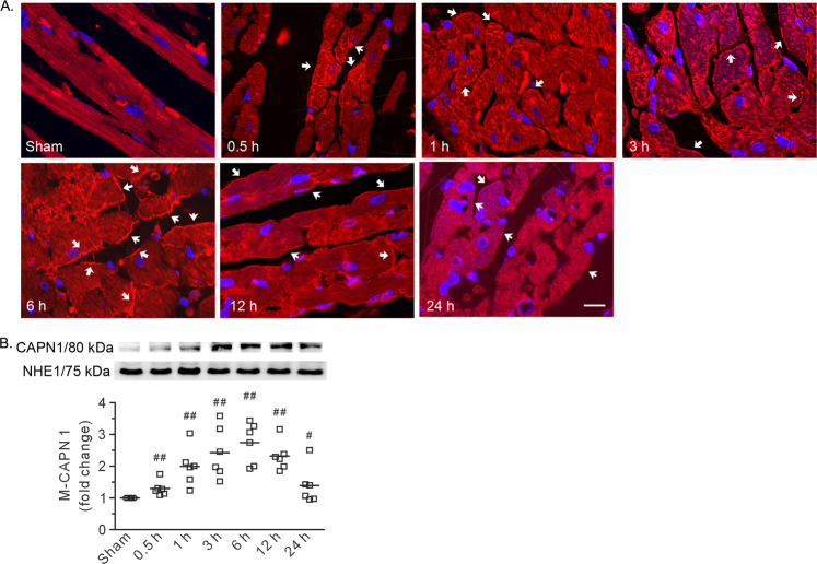
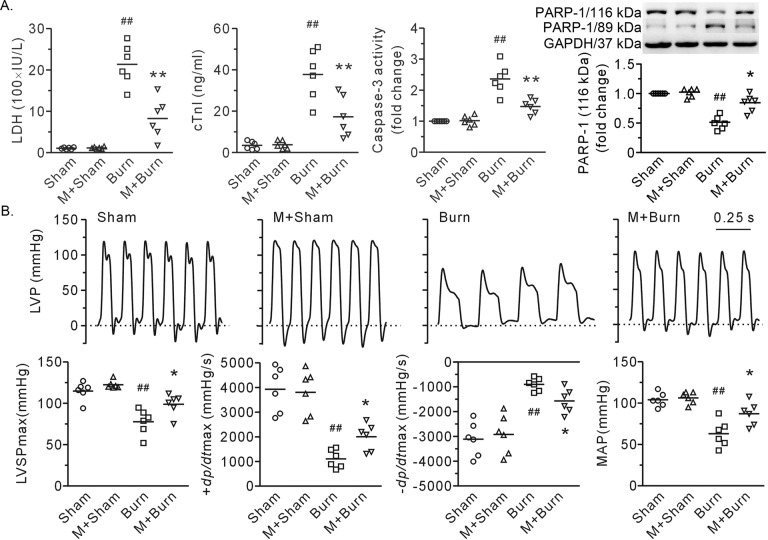
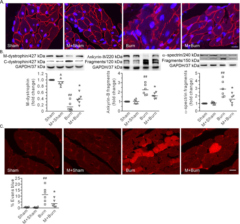
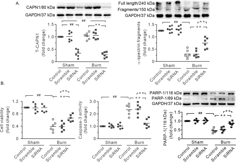
Stress cardiomyopathy is a major clinical complication after severe burn. Multiple upstream initiators have been identified; however, the downstream targets are not fully understood. This study assessed the role of the plasma membrane in this process and its relationship with the protease μ-calpain and tumor necrosis factor-alpha (TNF-α). Here, third-degree burn injury of approximately 40% of the total body surface area was established in rats. Plasma levels of LDH and cTnI and cardiac cell apoptosis increased at 0.5 h post burn, reached a peak at 6 h, and gradually declined at 24 h. This effect correlated well with not only the disruption of cytoskeletal proteins, including dystrophin and ankyrin-B, but also with the activation of μ-calpain, as indicated by the cleaved fragments of α-spectrin and membrane recruitment of the catalytic subunit CAPN1. More importantly, these alterations were diminished by blocking calpain activity with MDL28170. Burn injury markedly increased the cellular uptake of Evans blue, indicating membrane integrity disruption, and this effect was also reversed by MDL28170. Compared with those in the control group, cardiac cells in the burn plasma-treated group were more prone to damage, as indicated by a marked decrease in cell viability and increases in LDH release and apoptosis. Of note, these alterations were mitigated by CAPN1 siRNA. Moreover, after neutralizing TNF-α with rhTNFR:Fc, calpain activity was blocked, and heart function was improved. In conclusion, we identified μ-calpain as a trigger for severe burn-induced membrane disruption in the heart and provided evidence for the application of rhTNFR:Fc to inhibit calpain for cardioprotection.
应激性心肌病是严重烧伤后的一种主要临床并发症。已确定了多个上游启动因子;然而,下游靶点尚未完全明确。本研究评估了质膜在此过程中的作用及其与蛋白酶μ-钙蛋白酶和肿瘤坏死因子-α(TNF-α)的关系。在此,在大鼠中建立了约占全身表面积40%的三度烧伤损伤。烧伤后0.5小时,血浆中乳酸脱氢酶(LDH)和心肌肌钙蛋白I(cTnI)水平以及心肌细胞凋亡增加,在6小时达到峰值,并在24小时逐渐下降。这种效应不仅与包括肌营养不良蛋白和锚蛋白B在内的细胞骨架蛋白的破坏密切相关,而且与μ-钙蛋白酶的激活密切相关,α-血影蛋白的裂解片段和催化亚基钙蛋白酶1(CAPN1)的膜募集表明了这一点。更重要的是,用MDL28170阻断钙蛋白酶活性可减轻这些改变。烧伤损伤显著增加了伊文思蓝的细胞摄取,表明膜完整性受到破坏,MDL28170也可逆转这种效应。与对照组相比,烧伤血浆处理组的心肌细胞更容易受损,表现为细胞活力显著下降、LDH释放增加和凋亡增加。值得注意的是,CAPN1小干扰RNA(siRNA)可减轻这些改变。此外,用重组人肿瘤坏死因子受体:Fc(rhTNFR:Fc)中和TNF-α后,钙蛋白酶活性被阻断,心脏功能得到改善。总之,我们确定μ-钙蛋白酶是严重烧伤诱导心脏膜破坏的触发因素,并为应用rhTNFR:Fc抑制钙蛋白酶以保护心脏提供了证据。