Du Peng-Ran, Lu Hong-Ting, Lin Xi-Xiang, Wang Li-Feng, Wang Yan-Xia, Gu Xiao-Ming, Bai Xiao-Zhi, Tao Ke, Zhou Jing-Jun
1Department of Physiology and Pathophysiology, Fourth Military Medical University, No. 169 Changle West Road, Xi'an, 710032 China.
2Department of Biochemistry, Fourth Military Medical University, Xi'an, Shaanxi China.
Burns Trauma. 2018 Oct 8;6:28. doi: 10.1186/s41038-018-0130-3. eCollection 2018.
The molecular pattern of severe burn-induced acute lung injury, characterized by cell structure damage and leukocyte infiltration, remains unknown. This study aimed to determine whether calpain, a protease involved in both processes, mediates severe burn-induced acute lung injury.
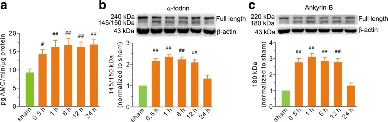
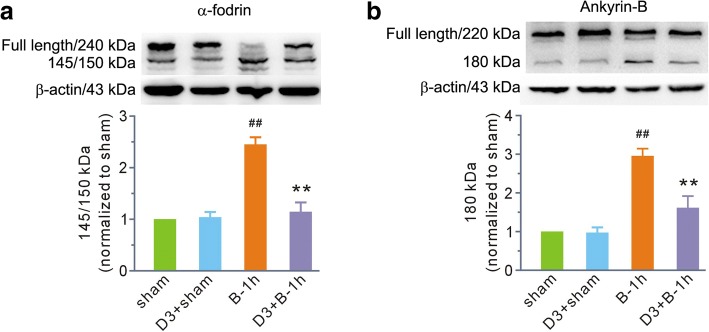
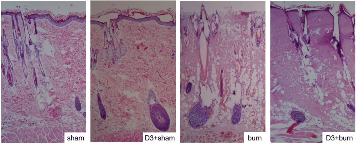
Rats received full-thickness scald burns covering 30% of the total body surface area, followed by instant fluid resuscitation. MDL28170 (Tocris Bioscience), an inhibitor of calpain, was given intravenously 1 h before or after the scald burn. The histological score, wet/dry weight ratio, and caspase-3 activity were examined to evaluate the degree of lung damage. Calpain activity and its source were detected by an assay kit and immunofluorescence staining. The proteolysis of membrane skeleton proteins α-fodrin and ankyrin-B, which are substrates of calpain, was measured by Western blot.
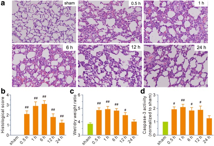
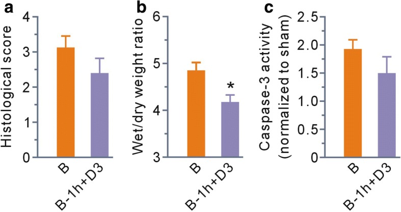
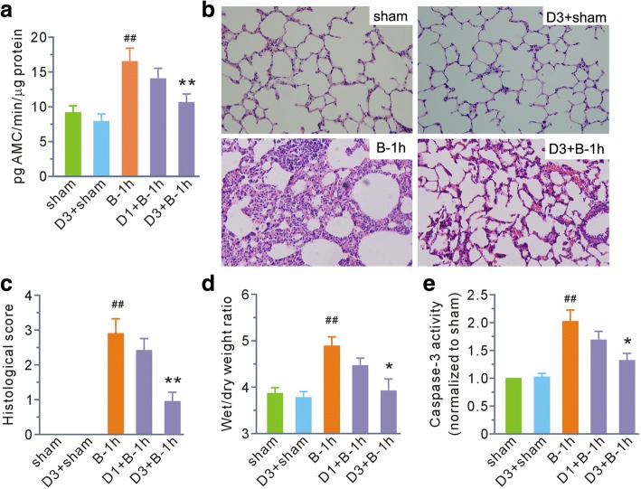
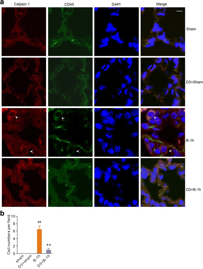
Time-course studies showed that tissue damage reached a peak between 1 and 6 h post-scald burn and gradually diminished at 24 h. More importantly, calpain activity reached peak levels at 1 h and was maintained until 24 h, paralleled by lung damage to some extent. Western blot showed that the levels of the proteolyzed forms of α-fodrin and ankyrin-B correlated well with the degree of damage. MDL28170 at a dose of 3 mg/kg b. w. given 1 h before burn injury not only antagonized the increase in calpain activity but also ameliorated scald burn-induced lung injury, including the degradation of α-fodrin and ankyrin-B. Immunofluorescence images revealed calpain 1 and CD45 double-positive cells in the lung tissue of rats exposed to scald burn injury, suggesting that leukocytes were a dominant source of calpain. Furthermore, this change was blocked by MDL28170. Finally, MDL28170 given at 1 h post-scald burn injury significantly ameliorated the wet/dry weight ratio compared with burn injury alone.
Calpain, a product of infiltrating leukocytes, is a mediator of scald burn-induced acute lung injury that involves enhancement of inflammation and proteolysis of membrane skeleton proteins. Its late effects warrant further study.
严重烧伤所致急性肺损伤的分子模式尚不清楚,其特征为细胞结构损伤和白细胞浸润。本研究旨在确定钙蛋白酶(一种参与上述两个过程的蛋白酶)是否介导严重烧伤所致急性肺损伤。
对大鼠进行占全身表面积30%的全层烫伤,随后立即进行液体复苏。在烫伤前或烫伤后1小时静脉注射钙蛋白酶抑制剂MDL28170(Tocris Bioscience公司)。通过检查组织学评分、湿/干重比和半胱天冬酶-3活性来评估肺损伤程度。用检测试剂盒和免疫荧光染色检测钙蛋白酶活性及其来源。通过蛋白质印迹法检测作为钙蛋白酶底物的膜骨架蛋白α-血影蛋白和锚蛋白B的蛋白水解情况。
时间进程研究表明,组织损伤在烫伤后1至6小时达到峰值,并在24小时逐渐减轻。更重要的是,钙蛋白酶活性在1小时达到峰值水平,并维持至24小时,在一定程度上与肺损伤平行。蛋白质印迹法显示,α-血影蛋白和锚蛋白B的蛋白水解形式水平与损伤程度密切相关。烧伤前1小时给予剂量为3mg/kg体重的MDL28170不仅能拮抗钙蛋白酶活性的增加,还能改善烫伤所致肺损伤,包括α-血影蛋白和锚蛋白B的降解。免疫荧光图像显示,烫伤大鼠肺组织中钙蛋白酶1和CD45双阳性细胞,提示白细胞是钙蛋白酶的主要来源。此外,这种变化被MDL28170阻断。最后,烫伤后1小时给予MDL28170与单纯烧伤相比,显著改善了湿/干重比。
钙蛋白酶作为浸润白细胞的产物,是烫伤所致急性肺损伤的介质,其涉及炎症增强和膜骨架蛋白的蛋白水解。其后期效应值得进一步研究。