Fang Kai, Kevil Christopher G
Inflammatory Bowel Disease Center, Vatche and Tamar Manoukian Division of Digestive Diseases, David Geffen School of Medicine, University of California, Los Angeles, California 90095, USA.
Department of Pathology, Louisiana State University Health Sciences Center, Shreveport, LA 71103, USA.
Int J Inflam. 2020 Mar 6;2020:6150942. doi: 10.1155/2020/6150942. eCollection 2020.
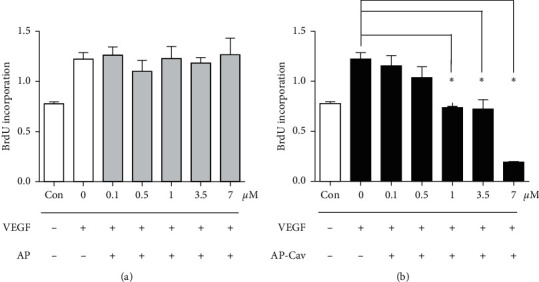
It has been reported that pathological angiogenesis contributes to both experimental colitis and inflammatory bowel disease. Recently, we demonstrated that endothelial caveolin-1 plays a key role in the pathological angiogenesis of dextran sodium sulfate (DSS) colitis. However, the molecular mechanism of caveolin-1 regulation of endothelial function is unknown. In this study, we examined how the antennapedia- (AP-) conjugated caveolin-1 scaffolding domain (AP-Cav) modulates vascular endothelial growth factor- (VEGF-) dependent colon endothelial cell angiogenic responses, as seen during colitis. We used mouse colon endothelial cells and found that AP-Cav significantly inhibited VEGF-mediated bromodeoxyuridine (BrdU) incorporation into colon microvascular endothelial cells. AP-Cav significantly blunted VEGF-dependent extracellular signal-regulated kinase 1/2 (ERK 1/2) phosphorylation at 10 minutes and 2 hours after stimulation, compared with the AP control peptide. AP-Cav + VEGF-A treatment also significantly increased c-Jun N-terminal kinase (JNK) phosphorylation at 2 hours. AP-Cav + VEGF-A treatment significantly downregulated retinoblastoma (Rb) protein levels, upregulated cleaved caspase-3 protein levels at 4 hours, and induced apoptosis. Thus, our study suggests that disruption of endothelial caveolin-1 function via the AP-Cav diverts VEGF signaling responses away from endothelial cell proliferation and toward apoptosis through the inhibition of mitogen-activated protein (MAP) kinase signaling and the induction of JNK-associated apoptosis.
据报道,病理性血管生成与实验性结肠炎和炎症性肠病均有关联。最近,我们证明内皮小窝蛋白-1在葡聚糖硫酸钠(DSS)结肠炎的病理性血管生成中起关键作用。然而,小窝蛋白-1调节内皮功能的分子机制尚不清楚。在本研究中,我们研究了与穿膜肽(AP)偶联的小窝蛋白-1支架结构域(AP-Cav)如何调节血管内皮生长因子(VEGF)依赖性结肠内皮细胞的血管生成反应,就像在结肠炎期间所观察到的那样。我们使用小鼠结肠内皮细胞,发现AP-Cav显著抑制VEGF介导的溴脱氧尿苷(BrdU)掺入结肠微血管内皮细胞。与AP对照肽相比,AP-Cav在刺激后10分钟和2小时显著减弱VEGF依赖性细胞外信号调节激酶1/2(ERK 1/2)的磷酸化。AP-Cav + VEGF-A处理在2小时时也显著增加c-Jun氨基末端激酶(JNK)的磷酸化。AP-Cav + VEGF-A处理显著下调视网膜母细胞瘤(Rb)蛋白水平,在4小时时上调裂解的半胱天冬酶-3蛋白水平,并诱导细胞凋亡。因此,我们的研究表明,通过AP-Cav破坏内皮小窝蛋白-1功能会使VEGF信号反应从内皮细胞增殖转向凋亡,这是通过抑制丝裂原活化蛋白(MAP)激酶信号传导和诱导JNK相关的细胞凋亡实现的。