• 文献检索
  • 文档翻译
  • 深度研究
  • 学术资讯
  • Suppr Zotero 插件Zotero 插件
  • 邀请有礼
  • 套餐&价格
  • 历史记录
应用&插件
Suppr Zotero 插件Zotero 插件浏览器插件Mac 客户端Windows 客户端微信小程序
定价
高级版会员购买积分包购买API积分包
服务
文献检索文档翻译深度研究API 文档MCP 服务
关于我们
关于 Suppr公司介绍联系我们用户协议隐私条款
关注我们

Suppr 超能文献

核心技术专利:CN118964589B侵权必究
粤ICP备2023148730 号-1Suppr @ 2026

文献检索

告别复杂PubMed语法,用中文像聊天一样搜索,搜遍4000万医学文献。AI智能推荐,让科研检索更轻松。

立即免费搜索

文件翻译

保留排版,准确专业,支持PDF/Word/PPT等文件格式,支持 12+语言互译。

免费翻译文档

深度研究

AI帮你快速写综述,25分钟生成高质量综述,智能提取关键信息,辅助科研写作。

立即免费体验

应用心脏扩散张量成像技术检测 ST 段抬高型心肌梗死患者的心肌内铁沉积。

Detection of Intramyocardial Iron in Patients Following ST-Elevation Myocardial Infarction Using Cardiac Diffusion Tensor Imaging.

机构信息

Biomedical Imaging Science Department, Leeds Institute of Cardiovascular and Metabolic Medicine, University of Leeds, Leeds Teaching Hospitals NHS Trust, Leeds, UK.

Institute for Biomedical Engineering, University and ETH Zurich, Zurich, Switzerland.

出版信息

J Magn Reson Imaging. 2022 Oct;56(4):1171-1181. doi: 10.1002/jmri.28063. Epub 2022 Jan 12.

DOI:10.1002/jmri.28063
PMID:35019174
原文链接:https://pmc.ncbi.nlm.nih.gov/articles/PMC9544509/
Abstract

BACKGROUND

Intramyocardial hemorrhage (IMH) following ST-elevation myocardial infarction (STEMI) is associated with poor prognosis. In cardiac magnetic resonance (MR), T2* mapping is the reference standard for detecting IMH while cardiac diffusion tensor imaging (cDTI) can characterize myocardial architecture via fractional anisotropy (FA) and mean diffusivity (MD) of water molecules. The value of cDTI in the detection of IMH is not currently known.

HYPOTHESIS

cDTI can detect IMH post-STEMI.

STUDY TYPE

Prospective.

SUBJECTS

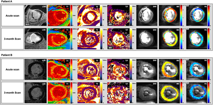
A total of 50 patients (20% female) scanned at 1-week (V1) and 3-month (V2) post-STEMI.

FIELD STRENGTH/SEQUENCE: A 3.0 T; inversion-recovery T1-weighted-imaging, multigradient-echo T2* mapping, spin-echo cDTI.

ASSESSMENT

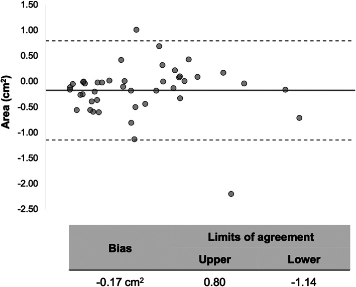
T2* maps were analyzed to detect IMH (defined as areas with T2* < 20 msec within areas of infarction). cDTI images were co-registered to produce averaged diffusion-weighted-images (DWIs), MD, and FA maps; hypointense areas were manually planimetered for IMH quantification.

STATISTICS

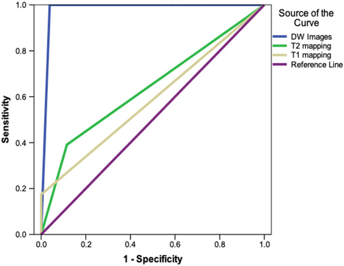
On averaged DWI, the presence of hypointense signal in areas matching IMH on T2* maps constituted to true-positive detection of iron. Independent samples t-tests were used to compare regional cDTI values. Results were considered statistically significant at P ≤ 0.05.

RESULTS

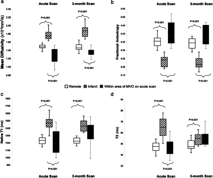
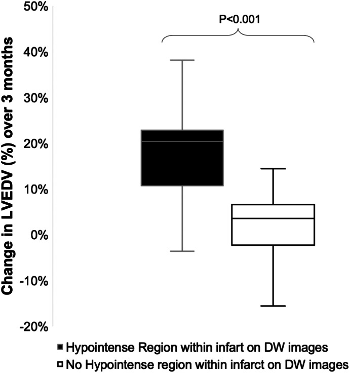
At V1, 24 patients had IMH on T2*. On averaged DWI, all 24 patients had hypointense signal in matching areas. IMH size derived using averaged-DWI was nonsignificantly greater than from T2* (2.0 ± 1.0 cm vs 1.89 ± 0.96 cm , P = 0.69). Compared to surrounding infarcted myocardium, MD was significantly reduced (1.29 ± 0.20 × 10  mm /sec vs 1.75 ± 0.16 × 10  mm /sec) and FA was significantly increased (0.40 ± 0.07 vs 0.23 ± 0.03) within areas of IMH. By V2, all 24 patients with acute IMH continued to have hypointense signals on averaged-DWI in the affected area. T2* detected IMH in 96% of these patients. Overall, averaged-DWI had 100% sensitivity and 96% specificity for the detection of IMH.

DATA CONCLUSION

This study demonstrates that the parameters MD and FA are susceptible to the paramagnetic properties of iron, enabling cDTI to detect IMH.

EVIDENCE LEVEL

1 TECHNICAL EFFICACY: Stage 2.

摘要

背景

ST 段抬高型心肌梗死(STEMI)后发生的心肌内出血(IMH)与预后不良有关。在心脏磁共振(CMR)中,T2* 映射是检测 IMH 的参考标准,而心脏扩散张量成像(cDTI)可以通过水分子的分数各向异性(FA)和平均扩散系数(MD)来描述心肌结构。cDTI 在检测 IMH 中的价值目前尚不清楚。

假设

cDTI 可以检测 STEMI 后的 IMH。

研究类型

前瞻性。

受试者

共 50 名患者(20%为女性),在 STEMI 后 1 周(V1)和 3 个月(V2)进行扫描。

磁场强度/序列:3.0T;反转恢复 T1 加权成像、多梯度回波 T2* 映射、自旋回波 cDTI。

评估

分析 T2* 图谱以检测 IMH(定义为在梗死区域内 T2* < 20msec 的区域)。将 cDTI 图像配准以生成平均扩散加权图像(DWI)、MD 和 FA 图;手动描绘低信号区域以进行 IMH 定量。

统计学

在平均 DWI 上,在与 T2* 图谱上 IMH 匹配的区域中出现低信号构成铁的真阳性检测。使用独立样本 t 检验比较区域 cDTI 值。结果 P ≤ 0.05 时认为具有统计学意义。

结果

在 V1 时,24 名患者的 T2* 上有 IMH。在平均 DWI 上,所有 24 名患者在匹配区域均有低信号。使用平均 DWI 得出的 IMH 大小与 T2* 无显著差异(2.0 ± 1.0cm 与 1.89 ± 0.96cm,P=0.69)。与周围梗死心肌相比,MD 明显降低(1.29 ± 0.20×10mm/sec 与 1.75 ± 0.16×10mm/sec),FA 明显增加(0.40 ± 0.07 与 0.23 ± 0.03)在 IMH 区域内。到 V2 时,所有 24 名急性 IMH 患者在受影响区域的平均 DWI 上继续有低信号。T2* 在这些患者中的 96%中检测到 IMH。总的来说,平均 DWI 对 IMH 的检测具有 100%的敏感性和 96%的特异性。

数据结论

本研究表明,参数 MD 和 FA 易受铁的顺磁性特性的影响,使 cDTI 能够检测 IMH。

证据水平

1 技术功效:2 级。

https://cdn.ncbi.nlm.nih.gov/pmc/blobs/4aff/9544509/4b464f506b6a/JMRI-56-1171-g002.jpg
https://cdn.ncbi.nlm.nih.gov/pmc/blobs/4aff/9544509/30bfa9a2e2fa/JMRI-56-1171-g004.jpg
https://cdn.ncbi.nlm.nih.gov/pmc/blobs/4aff/9544509/2b61d7b8dba0/JMRI-56-1171-g005.jpg
https://cdn.ncbi.nlm.nih.gov/pmc/blobs/4aff/9544509/1280aaa78c39/JMRI-56-1171-g001.jpg
https://cdn.ncbi.nlm.nih.gov/pmc/blobs/4aff/9544509/4268139bb6be/JMRI-56-1171-g003.jpg
https://cdn.ncbi.nlm.nih.gov/pmc/blobs/4aff/9544509/4b464f506b6a/JMRI-56-1171-g002.jpg

相似文献

1
Detection of Intramyocardial Iron in Patients Following ST-Elevation Myocardial Infarction Using Cardiac Diffusion Tensor Imaging.应用心脏扩散张量成像技术检测 ST 段抬高型心肌梗死患者的心肌内铁沉积。
J Magn Reson Imaging. 2022 Oct;56(4):1171-1181. doi: 10.1002/jmri.28063. Epub 2022 Jan 12.
2
Acute Microstructural Changes after ST-Segment Elevation Myocardial Infarction Assessed with Diffusion Tensor Imaging.急性 ST 段抬高型心肌梗死患者的磁共振弥散张量成像表现与急性微观结构变化的相关性研究。
Radiology. 2021 Apr;299(1):86-96. doi: 10.1148/radiol.2021203208. Epub 2021 Feb 9.
3
Head-to-head comparison of multiple cardiovascular magnetic resonance techniques for the detection and quantification of intramyocardial haemorrhage in patients with ST-elevation myocardial infarction.比较多种心血管磁共振技术对头检测和量化 ST 段抬高型心肌梗死患者心肌内出血的效果。
Eur Radiol. 2021 Mar;31(3):1245-1256. doi: 10.1007/s00330-020-07254-1. Epub 2020 Sep 14.
4
Diagnostic performance of T and T mapping to detect intramyocardial hemorrhage in reperfused ST-segment elevation myocardial infarction (STEMI) patients.T 及 T 映射技术诊断再灌注 ST 段抬高型心肌梗死(STEMI)患者心肌内出血的效能。
J Magn Reson Imaging. 2017 Sep;46(3):877-886. doi: 10.1002/jmri.25638. Epub 2017 Feb 15.
5
The relation between hypointense core, microvascular obstruction and intramyocardial haemorrhage in acute reperfused myocardial infarction assessed by cardiac magnetic resonance imaging.通过心脏磁共振成像评估急性再灌注心肌梗死中低信号核心、微血管阻塞与心肌内出血之间的关系。
Eur Radiol. 2014 Dec;24(12):3277-88. doi: 10.1007/s00330-014-3318-3. Epub 2014 Aug 6.
6
Residual Myocardial Iron Following Intramyocardial Hemorrhage During the Convalescent Phase of Reperfused ST-Segment-Elevation Myocardial Infarction and Adverse Left Ventricular Remodeling.再灌注ST段抬高型心肌梗死恢复期心肌内出血后残留心肌铁与左心室不良重构
Circ Cardiovasc Imaging. 2016 Oct;9(10):e004940. doi: 10.1161/CIRCIMAGING.116.004940.
7
Myocardial Extracellular Volume Fraction Allows Differentiation of Reversible Versus Irreversible Myocardial Damage and Prediction of Adverse Left Ventricular Remodeling of ST-Elevation Myocardial Infarction.心肌细胞外容积分数有助于鉴别可逆性与不可逆性心肌损伤,并预测ST段抬高型心肌梗死患者左心室不良重构。
J Magn Reson Imaging. 2020 Aug;52(2):476-487. doi: 10.1002/jmri.27047. Epub 2020 Jan 14.
8
Cardiac magnetic resonance imaging detection of intramyocardial hemorrhage in patients with ST-elevated myocardial infarction: comparison between susceptibility-weighted imaging and T1/T2 mapping techniques.心脏磁共振成像检测ST段抬高型心肌梗死患者心肌内出血:敏感性加权成像与T1/T2映射技术的比较
Quant Imaging Med Surg. 2024 Jan 3;14(1):476-488. doi: 10.21037/qims-23-591. Epub 2023 Nov 28.
9
Predictors of Intramyocardial Hemorrhage After Reperfused ST-Segment Elevation Myocardial Infarction.再灌注治疗的 ST 段抬高型心肌梗死患者心肌内出血的预测因素。
J Am Heart Assoc. 2017 Aug 15;6(8):e005651. doi: 10.1161/JAHA.117.005651.
10
The relationship between myocardial microstructure and strain in chronic infarction using cardiovascular magnetic resonance diffusion tensor imaging and feature tracking.应用心血管磁共振弥散张量成像和特征追踪技术研究慢性梗死心肌微结构与应变的关系。
J Cardiovasc Magn Reson. 2022 Nov 24;24(1):66. doi: 10.1186/s12968-022-00892-y.

引用本文的文献

1
Cardiac magnetic resonance imaging-derived pathophysiology and prognosis of diabetes mellitus with acute myocardial infarction after revascularization: a prospective cohort study.经心脏磁共振成像评估的糖尿病合并经血运重建治疗的急性心肌梗死后的病理生理学和预后:一项前瞻性队列研究。
Ann Med. 2024 Dec;56(1):2399751. doi: 10.1080/07853890.2024.2399751. Epub 2024 Sep 10.
2
The relationship between myocardial microstructure and strain in chronic infarction using cardiovascular magnetic resonance diffusion tensor imaging and feature tracking.应用心血管磁共振弥散张量成像和特征追踪技术研究慢性梗死心肌微结构与应变的关系。
J Cardiovasc Magn Reson. 2022 Nov 24;24(1):66. doi: 10.1186/s12968-022-00892-y.

本文引用的文献

1
Acute Microstructural Changes after ST-Segment Elevation Myocardial Infarction Assessed with Diffusion Tensor Imaging.急性 ST 段抬高型心肌梗死患者的磁共振弥散张量成像表现与急性微观结构变化的相关性研究。
Radiology. 2021 Apr;299(1):86-96. doi: 10.1148/radiol.2021203208. Epub 2021 Feb 9.
2
Hemorrhage promotes chronic adverse remodeling in acute myocardial infarction: a T , T and BOLD study.出血促进急性心肌梗死后的慢性不良重构:一项 T1、T2 和 BOLD 研究。
NMR Biomed. 2021 Jan;34(1):e4404. doi: 10.1002/nbm.4404. Epub 2020 Sep 1.
3
Insight Into Myocardial Microstructure of Athletes and Hypertrophic Cardiomyopathy Patients Using Diffusion Tensor Imaging.
利用扩散张量成像深入了解运动员和肥厚型心肌病患者的心肌微观结构
J Magn Reson Imaging. 2021 Jan;53(1):73-82. doi: 10.1002/jmri.27257. Epub 2020 Jun 18.
4
MRI of Reperfused Acute Myocardial Infarction Edema: ADC Quantification versus T1 and T2 Mapping.再灌注急性心肌梗死水肿的 MRI:ADC 定量与 T1 和 T2 映射。
Radiology. 2020 Jun;295(3):542-549. doi: 10.1148/radiol.2020192186. Epub 2020 Mar 24.
5
Influence of Myocardial Hemorrhage on Staging of Reperfused Myocardial Infarctions With T Cardiac Magnetic Resonance Imaging: Insights Into the Dependence on Infarction Type With Ex Vivo Validation.心肌出血对 T 心脏磁共振成像再灌注心肌梗死分期的影响:通过离体验证深入了解梗死类型的依赖性。
JACC Cardiovasc Imaging. 2019 Apr;12(4):693-703. doi: 10.1016/j.jcmg.2018.01.018. Epub 2018 Apr 18.
6
Feature-Tracking Global Longitudinal Strain Predicts Death in a Multicenter Population of Patients With Ischemic and Nonischemic Dilated Cardiomyopathy Incremental to Ejection Fraction and Late Gadolinium Enhancement.特征追踪整体纵向应变比射血分数和钆延迟增强更能预测缺血性和非缺血性扩张型心肌病患者的死亡:一项多中心研究。
JACC Cardiovasc Imaging. 2018 Oct;11(10):1419-1429. doi: 10.1016/j.jcmg.2017.10.024. Epub 2018 Jan 17.
7
An in-vivo comparison of stimulated-echo and motion compensated spin-echo sequences for 3 T diffusion tensor cardiovascular magnetic resonance at multiple cardiac phases.在 3T 扩散张量心血管磁共振的多个心动周期中,使用激发回波和运动补偿自旋回波序列的活体比较。
J Cardiovasc Magn Reson. 2018 Jan 3;20(1):1. doi: 10.1186/s12968-017-0425-8.
8
The impact of signal-to-noise ratio, diffusion-weighted directions and image resolution in cardiac diffusion tensor imaging - insights from the ex-vivo rat heart.信噪比、扩散加权方向和图像分辨率对心脏扩散张量成像的影响——来自离体大鼠心脏的观察。
J Cardiovasc Magn Reson. 2017 Nov 20;19(1):90. doi: 10.1186/s12968-017-0395-x.
9
Persistent Iron Within the Infarct Core After ST-Segment Elevation Myocardial Infarction: Implications for Left Ventricular Remodeling and Health Outcomes.ST 段抬高型心肌梗死梗死后梗死核心内持续存在的铁:对左心室重构和健康结局的影响。
JACC Cardiovasc Imaging. 2018 Sep;11(9):1248-1256. doi: 10.1016/j.jcmg.2017.08.027. Epub 2017 Nov 15.
10
Clinical recommendations for cardiovascular magnetic resonance mapping of T1, T2, T2* and extracellular volume: A consensus statement by the Society for Cardiovascular Magnetic Resonance (SCMR) endorsed by the European Association for Cardiovascular Imaging (EACVI).临床推荐意见:心血管磁共振 T1、T2、T2* 和细胞外容积mapping:心血管磁共振学会(SCMR)的共识声明,得到欧洲心血管影像协会(EACVI)的认可。
J Cardiovasc Magn Reson. 2017 Oct 9;19(1):75. doi: 10.1186/s12968-017-0389-8.