Department of Computer Science and Information Technology, University of Lahore, Lahore, Pakistan.
Department of Computer Science and Engineering, Sree Buddha College of Engineering, Alappuzha, Kerala, India.
J Healthc Eng. 2022 Jan 10;2022:2693621. doi: 10.1155/2022/2693621. eCollection 2022.
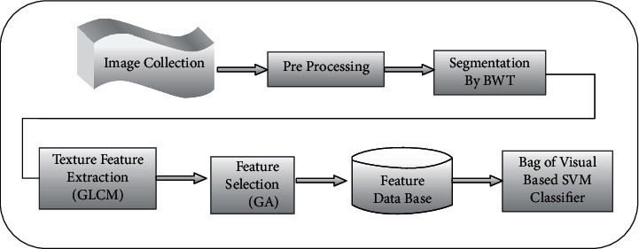
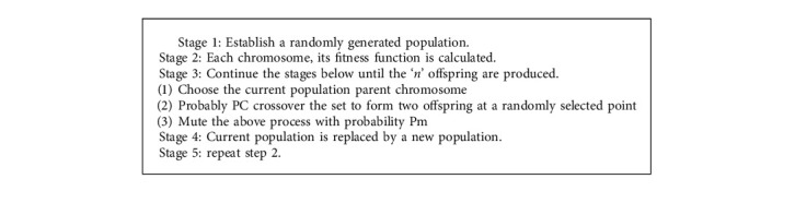
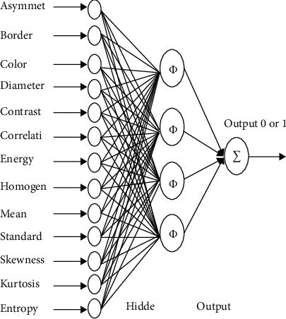
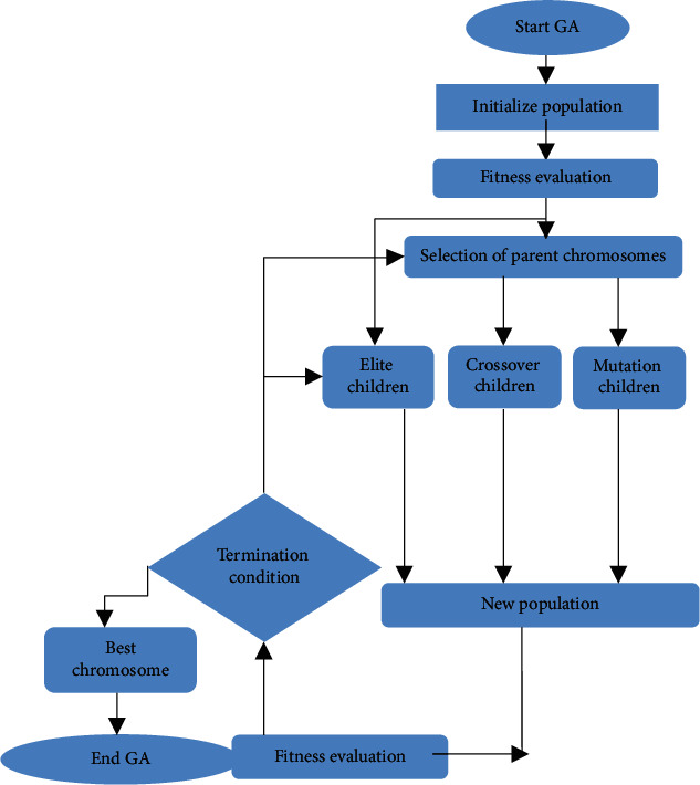
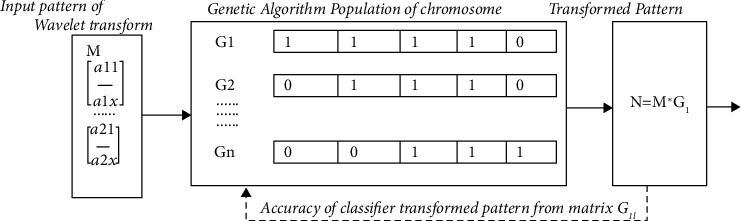
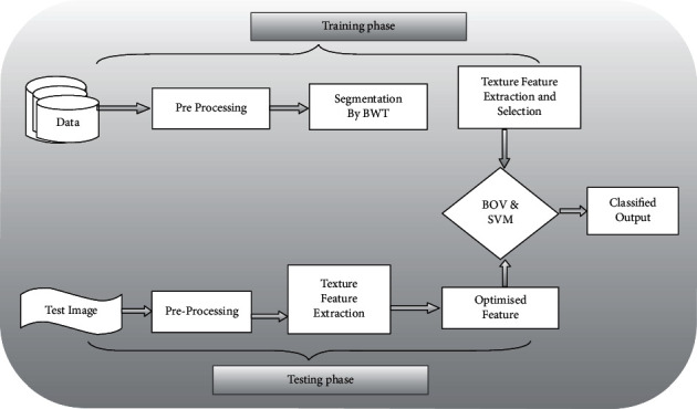
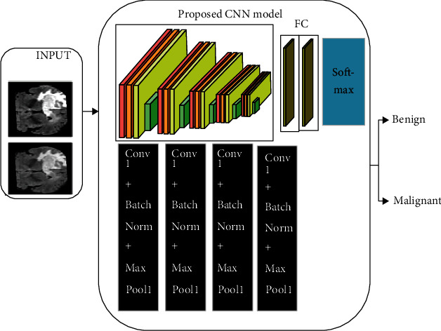
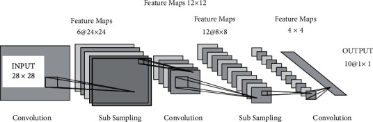
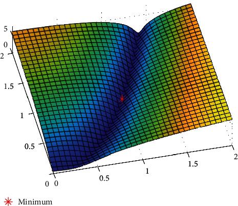
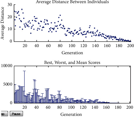
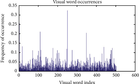
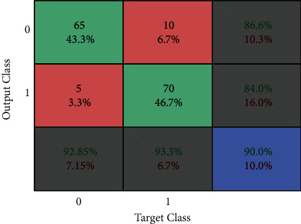
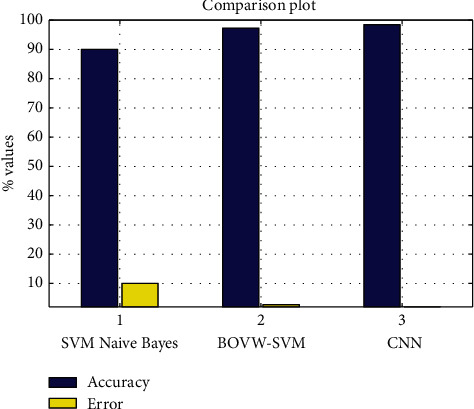
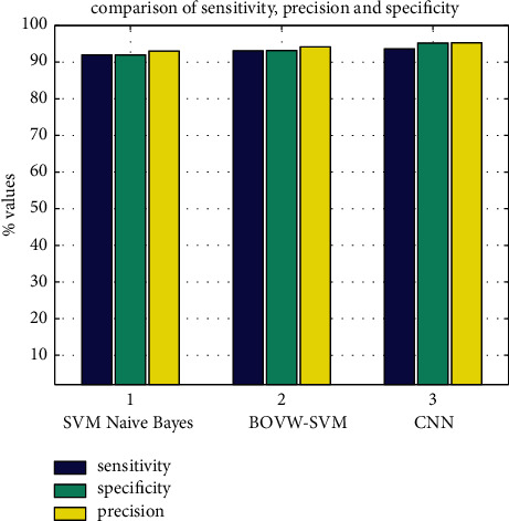
Radiology is a broad subject that needs more knowledge and understanding of medical science to identify tumors accurately. The need for a tumor detection program, thus, overcomes the lack of qualified radiologists. Using magnetic resonance imaging, biomedical image processing makes it easier to detect and locate brain tumors. In this study, a segmentation and detection method for brain tumors was developed using images from the MRI sequence as an input image to identify the tumor area. This process is difficult due to the wide variety of tumor tissues in the presence of different patients, and, in most cases, the similarity within normal tissues makes the task difficult. The main goal is to classify the brain in the presence of a brain tumor or a healthy brain. The proposed system has been researched based on Berkeley's wavelet transformation (BWT) and deep learning classifier to improve performance and simplify the process of medical image segmentation. Significant features are extracted from each segmented tissue using the gray-level-co-occurrence matrix (GLCM) method, followed by a feature optimization using a genetic algorithm. The innovative final result of the approach implemented was assessed based on accuracy, sensitivity, specificity, coefficient of dice, Jaccard's coefficient, spatial overlap, AVME, and FoM.
放射学是一个广泛的学科,需要更多的医学科学知识和理解才能准确识别肿瘤。因此,肿瘤检测程序的需求克服了合格放射科医生的缺乏。使用磁共振成像,生物医学图像处理使得更容易检测和定位脑肿瘤。在这项研究中,使用 MRI 序列的图像作为输入图像,开发了一种脑肿瘤的分割和检测方法,以识别肿瘤区域。由于存在不同患者的不同肿瘤组织的多样性,并且在大多数情况下,正常组织之间的相似性使得任务变得困难,因此这个过程很困难。主要目标是在存在脑肿瘤或健康大脑的情况下对大脑进行分类。所提出的系统是基于伯克利的小波变换(BWT)和深度学习分类器进行研究的,以提高性能并简化医学图像分割过程。使用灰度共生矩阵(GLCM)方法从每个分割组织中提取重要特征,然后使用遗传算法进行特征优化。基于准确性、敏感性、特异性、骰子系数、Jaccard 系数、空间重叠、AVME 和 FoM 评估实现的方法的创新最终结果。