Bahadure Nilesh Bhaskarrao, Ray Arun Kumar, Thethi Har Pal
School of Electronics Engineering, KIIT University, Bhubaneswar, Odisha, India.
Department of Electronics & Telecommunication Engineering, Lovely Professional University, Jalandhar, Punjab, India.
Int J Biomed Imaging. 2017;2017:9749108. doi: 10.1155/2017/9749108. Epub 2017 Mar 6.
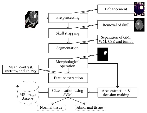
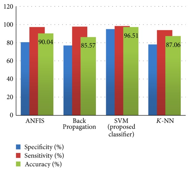
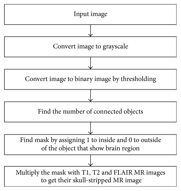
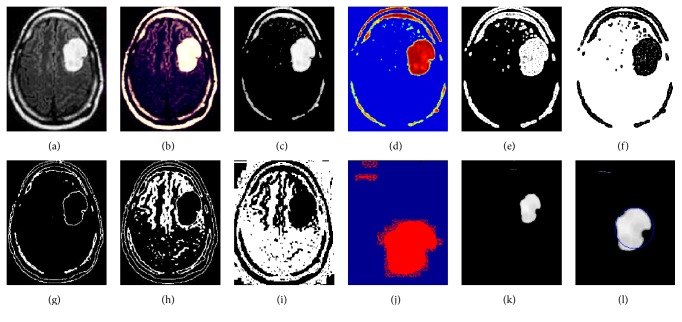
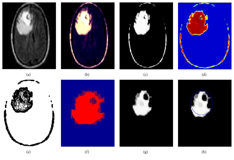
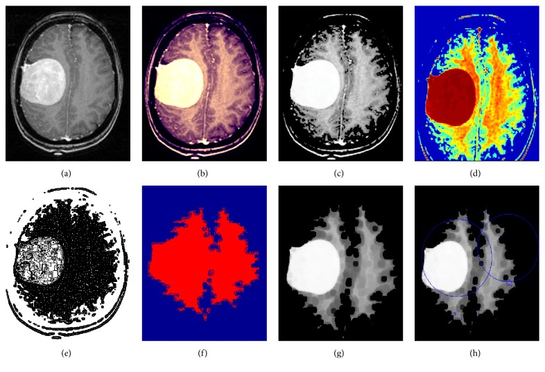
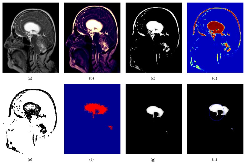
The segmentation, detection, and extraction of infected tumor area from magnetic resonance (MR) images are a primary concern but a tedious and time taking task performed by radiologists or clinical experts, and their accuracy depends on their experience only. So, the use of computer aided technology becomes very necessary to overcome these limitations. In this study, to improve the performance and reduce the complexity involves in the medical image segmentation process, we have investigated Berkeley wavelet transformation (BWT) based brain tumor segmentation. Furthermore, to improve the accuracy and quality rate of the support vector machine (SVM) based classifier, relevant features are extracted from each segmented tissue. The experimental results of proposed technique have been evaluated and validated for performance and quality analysis on magnetic resonance brain images, based on accuracy, sensitivity, specificity, and dice similarity index coefficient. The experimental results achieved 96.51% accuracy, 94.2% specificity, and 97.72% sensitivity, demonstrating the effectiveness of the proposed technique for identifying normal and abnormal tissues from brain MR images. The experimental results also obtained an average of 0.82 dice similarity index coefficient, which indicates better overlap between the automated (machines) extracted tumor region with manually extracted tumor region by radiologists. The simulation results prove the significance in terms of quality parameters and accuracy in comparison to state-of-the-art techniques.
从磁共振(MR)图像中分割、检测和提取受感染的肿瘤区域是一个主要关注点,但却是放射科医生或临床专家执行的一项繁琐且耗时的任务,其准确性仅取决于他们的经验。因此,使用计算机辅助技术变得非常必要,以克服这些限制。在本研究中,为了提高医学图像分割过程的性能并降低其复杂性,我们研究了基于伯克利小波变换(BWT)的脑肿瘤分割。此外,为了提高基于支持向量机(SVM)的分类器的准确性和质量率,从每个分割的组织中提取相关特征。基于准确性、敏感性、特异性和骰子相似性指数系数,对所提出技术的实验结果进行了评估和验证,以分析磁共振脑图像的性能和质量。实验结果达到了96.51%的准确率、94.2%的特异性和97.72%的敏感性,证明了所提出技术从脑MR图像中识别正常和异常组织的有效性。实验结果还获得了平均0.82的骰子相似性指数系数,这表明自动(机器)提取的肿瘤区域与放射科医生手动提取的肿瘤区域之间有更好的重叠。与现有技术相比,模拟结果证明了在质量参数和准确性方面的重要性。