Department of Biochemistry and Molecular Biology, State Key Laboratory for Zoonotic Diseases, School of Basic Medicine, Tongji Medical College, Huazhong University of Science and Technologygrid.33199.31, Wuhan, Hubei, China.
Institute of Health Inspection and Testing, Hubei Provincial Center for Disease Control and Prevention, Wuhan, Hubei, China.
mBio. 2021 Feb 22;13(1):e0316821. doi: 10.1128/mbio.03168-21. Epub 2022 Feb 1.
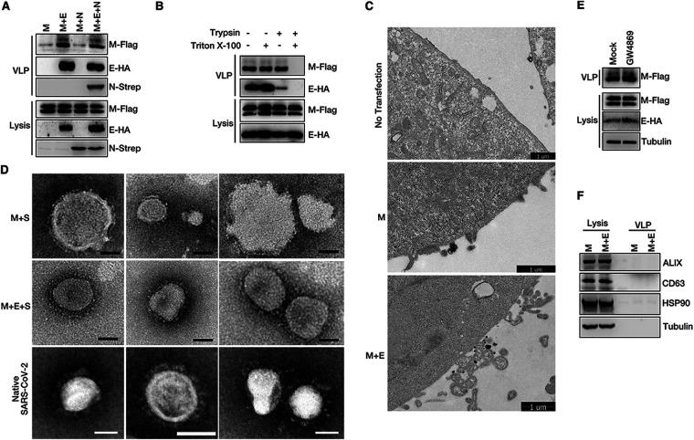
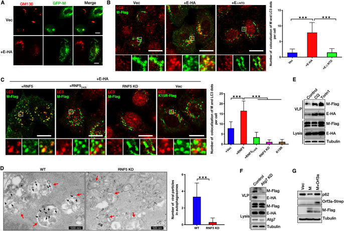
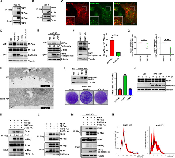
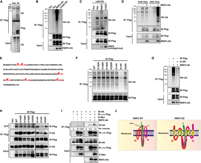
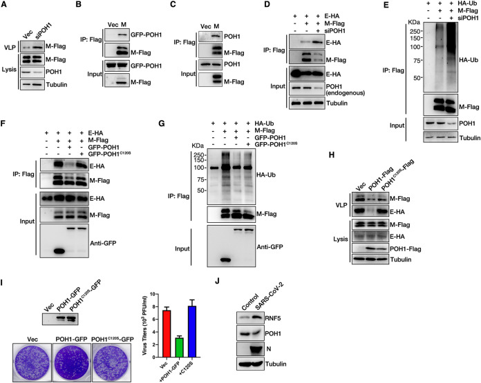
As an enveloped virus, severe acute respiratory syndrome coronavirus 2 (SARS-CoV-2) contains a membrane protein (M) that mediates viral release from cellular membranes. However, the molecular mechanisms of SARS-CoV-2 virion release remain poorly understood. In the present study, we performed RNA interference (RNAi) screening and identified the E3 ligase RNF5, which mediates the ubiquitination of SARS-CoV-2 M at residue K15 to enhance the interaction of the viral envelope protein (E) with M, whereas the deubiquitinating enzyme POH1 negatively regulates this process. The M-E complex ensures the uniform size of viral particles for viral maturation and mediates virion release. Moreover, M traffics from the Golgi apparatus to autophagosomes and uses autophagosomes for virion release, and this process is dependent on RNF5-mediated ubiquitin modification and M-E interaction. These results demonstrate that ubiquitin modification of SARS-CoV-2 M stabilizes the M-E complex and uses autophagosomes for virion release. Enveloped virus particles are released from the membranes of host cells, and viral membrane proteins (M) are critical for this process. A better understanding of the molecular mechanisms of SARS-CoV-2 assembly and budding is critical for the development of antiviral therapies. Envelope protein (E) and M of SARS-CoV-2 form complexes to mediate viral assembly and budding. RNF5 was identified to play a role as the E3 ligase, and POH1 was demonstrated to function as the deubiquitinating enzyme of SARS-CoV-2 M. The two components collectively regulate the interaction of M with E to promote viral assembly and budding. Ubiquitinated M uses autophagosomes for viral release. Our findings provide insights into the mechanisms of SARS-CoV-2 assembly and budding, demonstrating the importance of ubiquitination modification and autophagy in viral replication.
作为一种包膜病毒,严重急性呼吸综合征冠状病毒 2(SARS-CoV-2)包含一种膜蛋白(M),介导病毒从细胞膜释放。然而,SARS-CoV-2 病毒粒子释放的分子机制仍知之甚少。在本研究中,我们进行了 RNA 干扰(RNAi)筛选,鉴定出 E3 连接酶 RNF5,它介导 SARS-CoV-2 M 残基 K15 的泛素化,增强病毒包膜蛋白(E)与 M 的相互作用,而去泛素化酶 POH1 则负调控这一过程。M-E 复合物确保病毒颗粒大小均匀,有利于病毒成熟,并介导病毒粒子释放。此外,M 从高尔基体运输到自噬体,并利用自噬体进行病毒粒子释放,这一过程依赖于 RNF5 介导的泛素修饰和 M-E 相互作用。这些结果表明,SARS-CoV-2 M 的泛素修饰稳定了 M-E 复合物,并利用自噬体进行病毒粒子释放。 包膜病毒粒子从宿主细胞的膜中释放出来,病毒膜蛋白(M)对于这一过程至关重要。更好地理解 SARS-CoV-2 的组装和出芽的分子机制对于开发抗病毒疗法至关重要。SARS-CoV-2 的包膜蛋白(E)和 M 形成复合物,介导病毒的组装和出芽。鉴定出 RNF5 作为 E3 连接酶发挥作用,POH1 被证明作为 SARS-CoV-2 M 的去泛素化酶发挥作用。这两个组件共同调节 M 与 E 的相互作用,促进病毒的组装和出芽。泛素化的 M 利用自噬体进行病毒释放。我们的研究结果提供了 SARS-CoV-2 组装和出芽机制的深入了解,证明了泛素化修饰和自噬在病毒复制中的重要性。