Smeeton Joanna, Natarajan Natasha, Anderson Troy, Tseng Kuo-Chang, Fabian Peter, Crump J Gage
Department of Rehabilitation and Regenerative Medicine, Columbia Stem Cell Initiative, Columbia University Irving Medical Center, Columbia University, New York, NY, United States.
Department of Genetics and Development, Columbia Stem Cell Initiative, Columbia University Irving Medical Center, Columbia University, New York, NY, United States.
Front Cell Dev Biol. 2022 Jan 20;9:777787. doi: 10.3389/fcell.2021.777787. eCollection 2021.
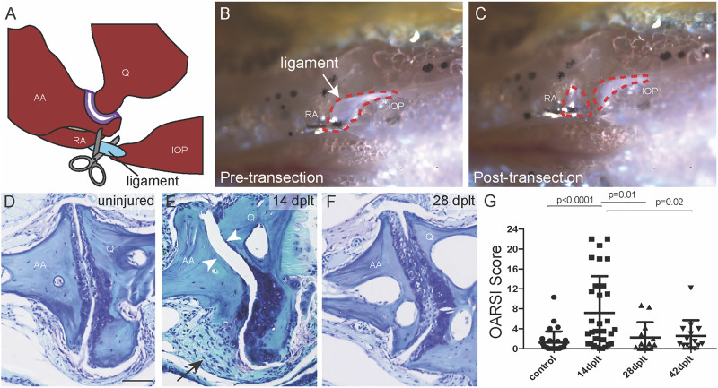
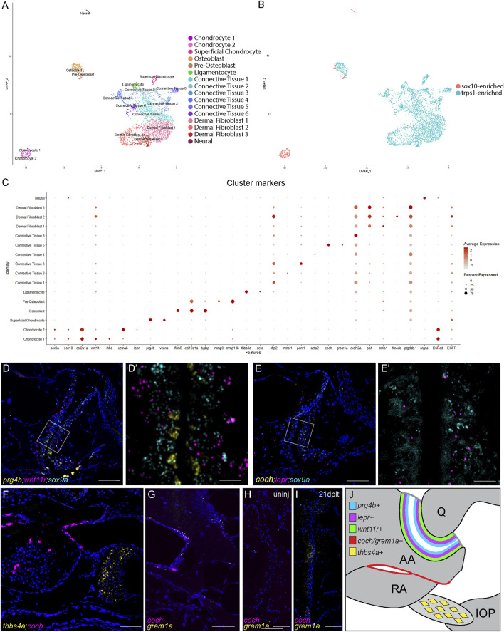
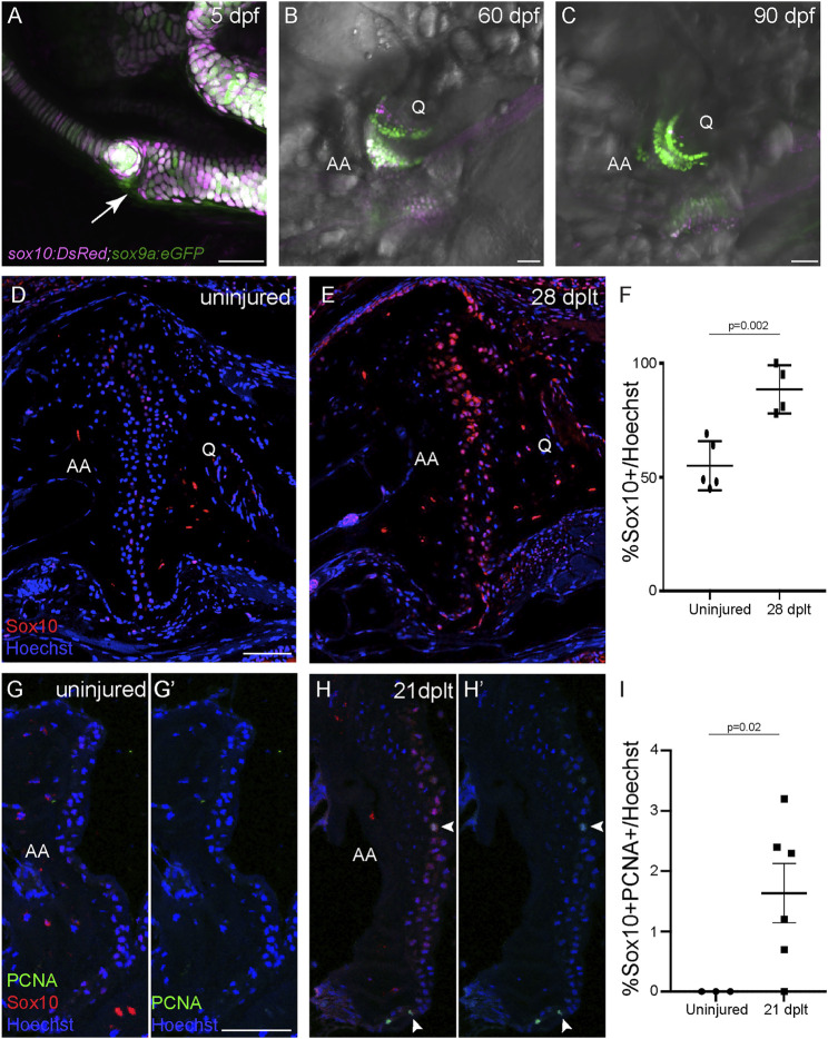
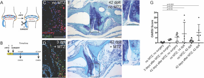
The poor intrinsic repair capacity of mammalian joint cartilage likely contributes to the high incidence of arthritis worldwide. Adult zebrafish can regenerate many structures that show limited or no healing capacity in mammals, including the jawbone. To test whether zebrafish can also regenerate damaged joints, we developed a surgical injury model in which the zebrafish jaw joint is destabilized transection of the major jaw joint ligament, the interopercular-mandibular (IOM). Unilateral transection of the IOM ligament in 1-year-old fish resulted in an initial reduction of jaw joint cartilage by 14 days, with full regeneration of joint cartilage by 28 days. Joint cartilage regeneration involves the re-entry of articular chondrocytes into the cell cycle and the upregulated expression of , a marker of developing chondrocytes in the embryo that becomes restricted to a subset of joint chondrocytes in adults. Genetic ablation of these -expressing chondrocytes shows that they are essential for joint cartilage regeneration. To uncover the potential source of new chondrocytes during joint regeneration, we performed single-cell RNA sequencing of the uninjured adult jaw joint and identified multiple skeletal, connective tissue, and fibroblast subtypes. In particular, we uncovered a joint-specific periosteal population expressing and , with the jaw joint chondrocytes marked by expression during regeneration. Our findings demonstrate the capacity of zebrafish to regenerate adult joint cartilage and identify candidate cell types that can be tested for their roles in regenerative response.
哺乳动物关节软骨固有的修复能力较差,这可能是全球关节炎高发的原因之一。成年斑马鱼能够再生许多在哺乳动物中愈合能力有限或无法愈合的结构,包括颌骨。为了测试斑马鱼是否也能再生受损关节,我们开发了一种手术损伤模型,通过切断主要的颌关节韧带——鳃盖骨 - 下颌骨(IOM),使斑马鱼的颌关节失稳。对1岁的斑马鱼单侧切断IOM韧带,导致颌关节软骨在14天时初步减少,到28天时关节软骨完全再生。关节软骨再生涉及关节软骨细胞重新进入细胞周期以及 的表达上调, 是胚胎中正在发育的软骨细胞的标志物,在成体中仅限于一部分关节软骨细胞表达。对这些表达 的软骨细胞进行基因消融表明,它们对关节软骨再生至关重要。为了揭示关节再生过程中新软骨细胞的潜在来源,我们对未受伤的成年颌关节进行了单细胞RNA测序,并鉴定出多种骨骼、结缔组织和成纤维细胞亚型。特别是,我们发现了一个关节特异性的骨膜群体,其表达 和 ,在再生过程中以 的表达为标记的颌关节软骨细胞。我们的研究结果证明了斑马鱼再生成年关节软骨的能力,并确定了可测试其在再生反应中作用的候选细胞类型。