Cardiac Arrhythmia Center and Neurocardiology Research Program of Excellence, David Geffen School of Medicine, University of California, Los Angeles, Los Angeles, California, USA.
Department of Biomedical Engineering, George Washington University, Washington, DC, USA.
JCI Insight. 2022 Feb 8;7(3):e153913. doi: 10.1172/jci.insight.153913.
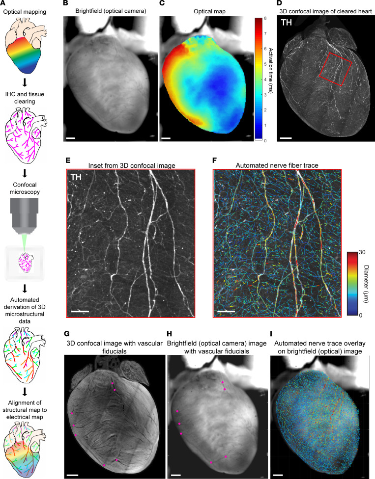
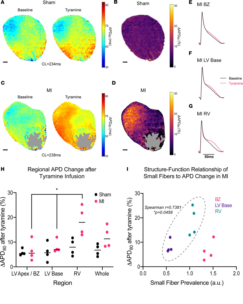
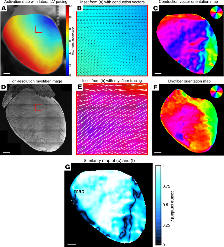
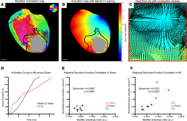
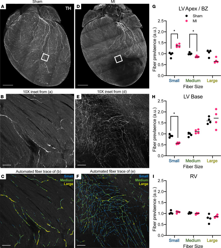
Remodeling of injured sympathetic nerves on the heart after myocardial infarction (MI) contributes to adverse outcomes such as sudden arrhythmic death, yet the underlying structural mechanisms are poorly understood. We sought to examine microstructural changes on the heart after MI and to directly link these changes with electrical dysfunction. We developed a high-resolution pipeline for anatomically precise alignment of electrical maps with structural myofiber and nerve-fiber maps created by customized computer vision algorithms. Using this integrative approach in a mouse model, we identified distinct structure-function correlates to objectively delineate the infarct border zone, a known source of arrhythmias after MI. During tyramine-induced sympathetic nerve activation, we demonstrated regional patterns of altered electrical conduction aligned directly with altered neuroeffector junction distribution, pointing to potential neural substrates for cardiac arrhythmia. This study establishes a synergistic framework for examining structure-function relationships after MI with microscopic precision that has potential to advance understanding of arrhythmogenic mechanisms.
心肌梗死后(MI)受伤的交感神经重塑导致心律失常性猝死等不良后果,但潜在的结构机制尚不清楚。我们试图检查 MI 后心脏的微观结构变化,并将这些变化与电功能障碍直接联系起来。我们开发了一种高分辨率的管道,用于通过定制的计算机视觉算法创建的电图谱与结构肌纤维和神经纤维图谱进行解剖精确对准。在小鼠模型中使用这种综合方法,我们确定了不同的结构-功能相关性,以客观地区分梗死边界区,这是 MI 后心律失常的已知来源。在去甲肾上腺素诱导的交感神经激活期间,我们证明了电传导的区域模式与神经效应器连接分布的改变直接一致,这表明了心律失常的潜在神经底物。这项研究建立了一个具有微观精度的 MI 后检查结构-功能关系的协同框架,具有推进心律失常机制理解的潜力。