• 文献检索
  • 文档翻译
  • 深度研究
  • 学术资讯
  • Suppr Zotero 插件Zotero 插件
  • 邀请有礼
  • 套餐&价格
  • 历史记录
应用&插件
Suppr Zotero 插件Zotero 插件浏览器插件Mac 客户端Windows 客户端微信小程序
定价
高级版会员购买积分包购买API积分包
服务
文献检索文档翻译深度研究API 文档MCP 服务
关于我们
关于 Suppr公司介绍联系我们用户协议隐私条款
关注我们

Suppr 超能文献

核心技术专利:CN118964589B侵权必究
粤ICP备2023148730 号-1Suppr @ 2026

文献检索

告别复杂PubMed语法,用中文像聊天一样搜索,搜遍4000万医学文献。AI智能推荐,让科研检索更轻松。

立即免费搜索

文件翻译

保留排版,准确专业,支持PDF/Word/PPT等文件格式,支持 12+语言互译。

免费翻译文档

深度研究

AI帮你快速写综述,25分钟生成高质量综述,智能提取关键信息,辅助科研写作。

立即免费体验

肠道上皮内离散的上皮囊中,细菌微生物群诱导的固有干扰素 - lambda 反应刺激了抢先的抗病毒防御。

Homeostatic interferon-lambda response to bacterial microbiota stimulates preemptive antiviral defense within discrete pockets of intestinal epithelium.

机构信息

Department of Molecular Microbiology and Immunology, Oregon Health & Science University, Portland, United States.

Department of Medicine, Washington University School of Medicine, St. Louis, United States.

出版信息

Elife. 2022 Feb 9;11:e74072. doi: 10.7554/eLife.74072.

DOI:10.7554/eLife.74072
PMID:35137688
原文链接:https://pmc.ncbi.nlm.nih.gov/articles/PMC8853662/
Abstract

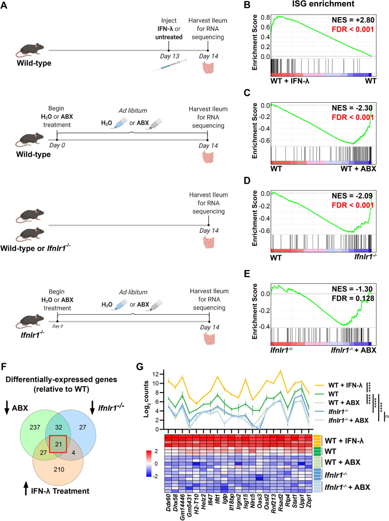
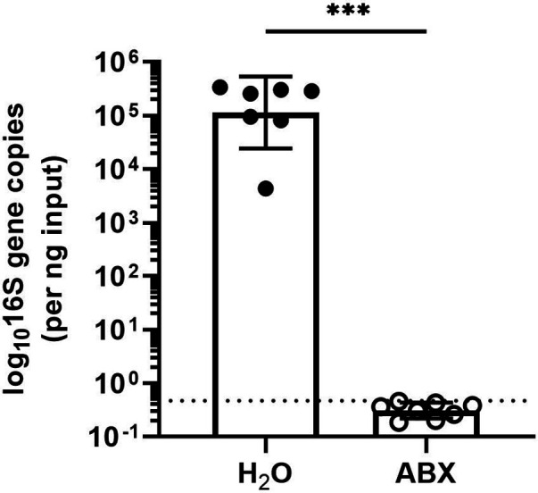
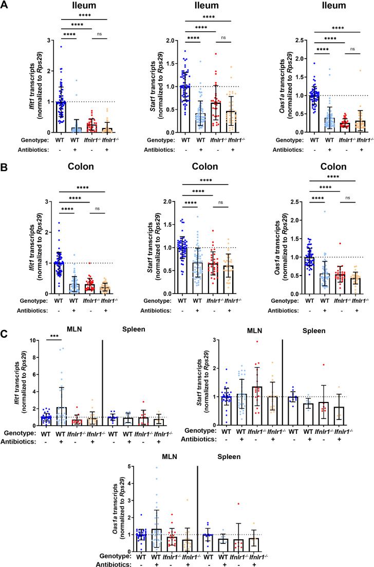
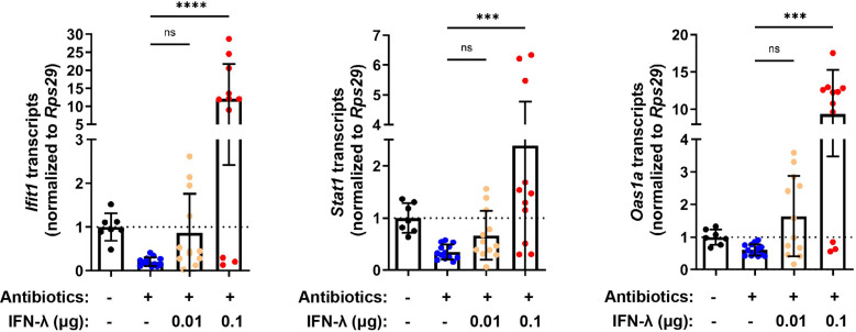
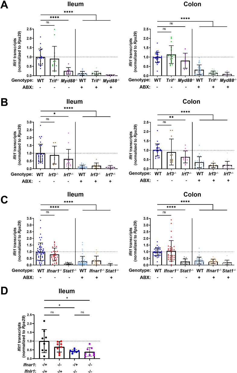
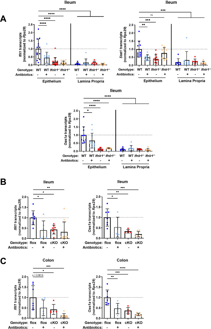
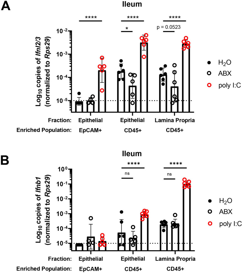
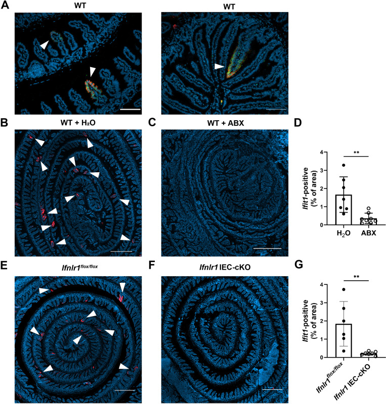
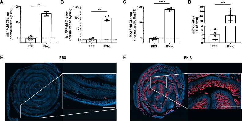
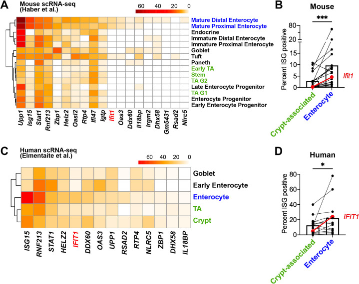
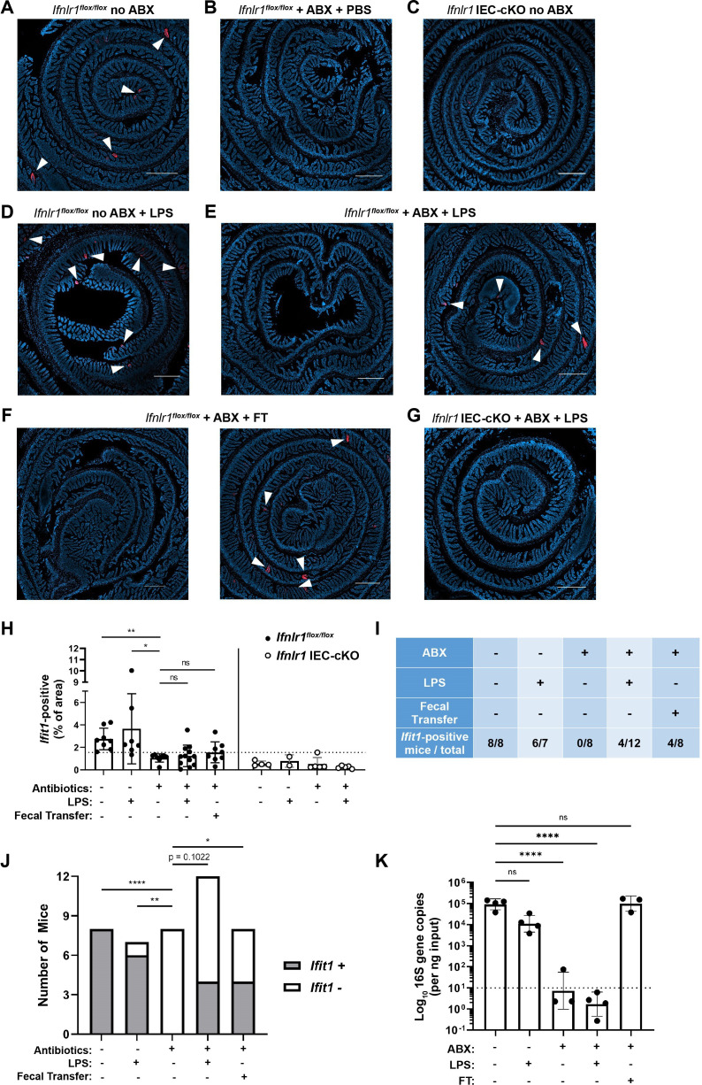
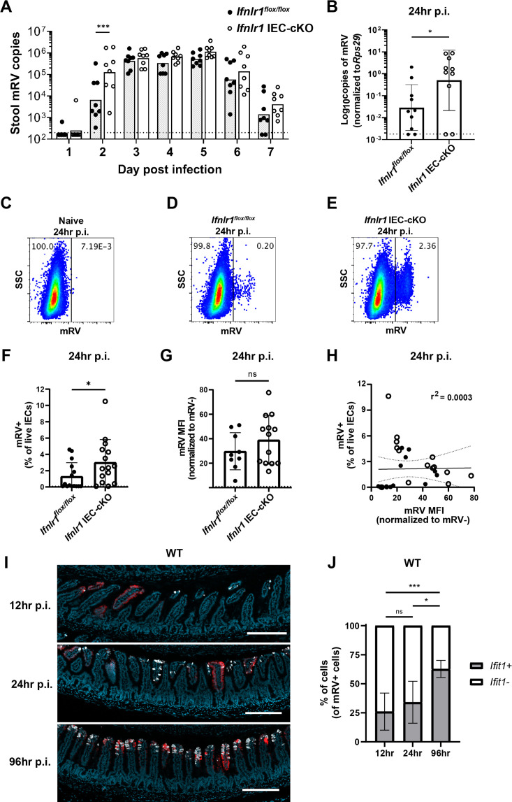
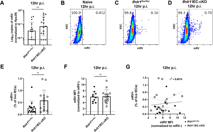
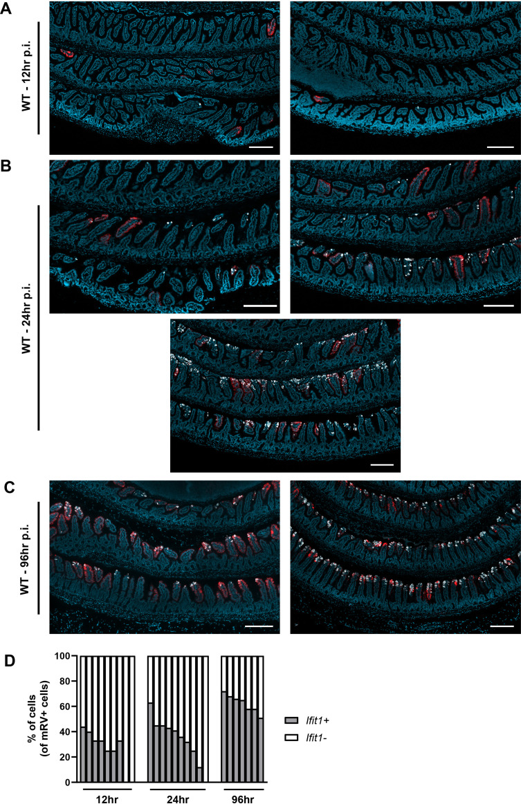
Interferon-lambda (IFN-λ) protects intestinal epithelial cells (IECs) from enteric viruses by inducing expression of antiviral IFN-stimulated genes (ISGs). Here, we find that bacterial microbiota stimulate a homeostatic ISG signature in the intestine of specific pathogen-free mice. This homeostatic ISG expression is restricted to IECs, depends on IEC-intrinsic expression of IFN-λ receptor (), and is associated with IFN-λ production by leukocytes. Strikingly, imaging of these homeostatic ISGs reveals localization to pockets of the epithelium and concentration in mature IECs. Correspondingly, a minority of mature IECs express these ISGs in public single-cell RNA sequencing datasets from mice and humans. Furthermore, we assessed the ability of orally administered bacterial components to restore localized ISGs in mice lacking bacterial microbiota. Lastly, we find that IECs lacking are hyper-susceptible to initiation of murine rotavirus infection. These observations indicate that bacterial microbiota stimulate ISGs in localized regions of the intestinal epithelium at homeostasis, thereby preemptively activating antiviral defenses in vulnerable IECs to improve host defense against enteric viruses.

摘要

干扰素-λ(IFN-λ)通过诱导抗病毒干扰素刺激基因(ISGs)的表达来保护肠上皮细胞(IECs)免受肠病毒感染。在这里,我们发现细菌微生物群会刺激无特定病原体小鼠肠道中的稳态 ISG 特征。这种稳态 ISG 表达仅限于 IECs,依赖于 IEC 固有表达的干扰素-λ受体(),并与白细胞产生 IFN-λ 有关。引人注目的是,对这些稳态 ISGs 的成像显示它们定位于上皮细胞的口袋中,并集中在成熟的 IECs 中。相应地,在来自小鼠和人类的公共单细胞 RNA 测序数据集中,少数成熟的 IEC 表达这些 ISGs。此外,我们评估了口服给予细菌成分在缺乏细菌微生物群的小鼠中恢复局部 ISGs 的能力。最后,我们发现缺乏的 IECs 对启动鼠轮状病毒感染非常敏感。这些观察结果表明,细菌微生物群在肠道上皮的局部区域刺激 ISGs,从而在脆弱的 IECs 中预先激活抗病毒防御,以改善宿主对肠道病毒的防御。

https://cdn.ncbi.nlm.nih.gov/pmc/blobs/ebc2/8853662/d150024dffff/elife-74072-sa2-fig1.jpg
https://cdn.ncbi.nlm.nih.gov/pmc/blobs/ebc2/8853662/c1e7a0debdcf/elife-74072-fig1.jpg
https://cdn.ncbi.nlm.nih.gov/pmc/blobs/ebc2/8853662/bf0e427e2fa4/elife-74072-fig1-figsupp1.jpg
https://cdn.ncbi.nlm.nih.gov/pmc/blobs/ebc2/8853662/4183fcc3aa74/elife-74072-fig1-figsupp2.jpg
https://cdn.ncbi.nlm.nih.gov/pmc/blobs/ebc2/8853662/ec423b9253fa/elife-74072-fig1-figsupp3.jpg
https://cdn.ncbi.nlm.nih.gov/pmc/blobs/ebc2/8853662/a67cf21fd19e/elife-74072-fig2.jpg
https://cdn.ncbi.nlm.nih.gov/pmc/blobs/ebc2/8853662/756b49f1cd2b/elife-74072-fig2-figsupp1.jpg
https://cdn.ncbi.nlm.nih.gov/pmc/blobs/ebc2/8853662/bfa5f5c49964/elife-74072-fig2-figsupp2.jpg
https://cdn.ncbi.nlm.nih.gov/pmc/blobs/ebc2/8853662/551656621e12/elife-74072-fig3.jpg
https://cdn.ncbi.nlm.nih.gov/pmc/blobs/ebc2/8853662/f20fba5703a2/elife-74072-fig4.jpg
https://cdn.ncbi.nlm.nih.gov/pmc/blobs/ebc2/8853662/1f9f8981d476/elife-74072-fig5.jpg
https://cdn.ncbi.nlm.nih.gov/pmc/blobs/ebc2/8853662/a66276a616b8/elife-74072-fig5-figsupp1.jpg
https://cdn.ncbi.nlm.nih.gov/pmc/blobs/ebc2/8853662/f685bedd3eec/elife-74072-fig6.jpg
https://cdn.ncbi.nlm.nih.gov/pmc/blobs/ebc2/8853662/94d65b878a4e/elife-74072-fig6-figsupp1.jpg
https://cdn.ncbi.nlm.nih.gov/pmc/blobs/ebc2/8853662/f94525270790/elife-74072-fig6-figsupp2.jpg
https://cdn.ncbi.nlm.nih.gov/pmc/blobs/ebc2/8853662/376b83e498b3/elife-74072-fig7.jpg
https://cdn.ncbi.nlm.nih.gov/pmc/blobs/ebc2/8853662/7d5bd8df5643/elife-74072-fig8.jpg
https://cdn.ncbi.nlm.nih.gov/pmc/blobs/ebc2/8853662/04acdb95c581/elife-74072-fig8-figsupp1.jpg
https://cdn.ncbi.nlm.nih.gov/pmc/blobs/ebc2/8853662/a186d088a7c0/elife-74072-fig9.jpg
https://cdn.ncbi.nlm.nih.gov/pmc/blobs/ebc2/8853662/b330d56abb0a/elife-74072-fig9-figsupp1.jpg
https://cdn.ncbi.nlm.nih.gov/pmc/blobs/ebc2/8853662/12dbdd578e6d/elife-74072-fig9-figsupp2.jpg
https://cdn.ncbi.nlm.nih.gov/pmc/blobs/ebc2/8853662/eefc22ba91e1/elife-74072-fig9-figsupp3.jpg
https://cdn.ncbi.nlm.nih.gov/pmc/blobs/ebc2/8853662/d150024dffff/elife-74072-sa2-fig1.jpg

相似文献

1
Homeostatic interferon-lambda response to bacterial microbiota stimulates preemptive antiviral defense within discrete pockets of intestinal epithelium.肠道上皮内离散的上皮囊中,细菌微生物群诱导的固有干扰素 - lambda 反应刺激了抢先的抗病毒防御。
Elife. 2022 Feb 9;11:e74072. doi: 10.7554/eLife.74072.
2
Expression of on Intestinal Epithelial Cells Is Critical to the Antiviral Effects of Interferon Lambda against Norovirus and Reovirus.[具体蛋白名称]在肠道上皮细胞上的表达对于干扰素λ对诺如病毒和呼肠孤病毒的抗病毒作用至关重要。 (注:原文中“Expression of on”缺少具体蛋白名称,这里补充了[具体蛋白名称]以便译文完整通顺)
J Virol. 2017 Mar 13;91(7). doi: 10.1128/JVI.02079-16. Print 2017 Apr 1.
3
Selective Interferon Responses of Intestinal Epithelial Cells Minimize Tumor Necrosis Factor Alpha Cytotoxicity.肠上皮细胞的选择性干扰素反应可最大程度地降低肿瘤坏死因子 α 的细胞毒性。
J Virol. 2020 Oct 14;94(21). doi: 10.1128/JVI.00603-20.
4
The TLR3/IRF1/Type III IFN Axis Facilitates Antiviral Responses against Enterovirus Infections in the Intestine.TLR3/IRF1/III 型 IFN 轴促进肠道对抗肠道病毒感染的抗病毒反应。
mBio. 2020 Nov 17;11(6):e02540-20. doi: 10.1128/mBio.02540-20.
5
Intestinal antiviral signaling is controlled by autophagy gene independent of the microbiota.肠道抗病毒信号传导由自噬基因控制,与微生物群无关。
Autophagy. 2022 May;18(5):1062-1077. doi: 10.1080/15548627.2021.1968607. Epub 2021 Sep 14.
6
Rotavirus Degrades Multiple Interferon (IFN) Type Receptors To Inhibit IFN Signaling and Protects against Mortality from Endotoxin in Suckling Mice.轮状病毒降解多种干扰素 (IFN) 型受体以抑制 IFN 信号转导并保护乳鼠免受内毒素引起的死亡。
J Virol. 2017 Dec 14;92(1). doi: 10.1128/JVI.01394-17. Print 2018 Jan 1.
7
Rotavirus Reprograms Multiple Interferon Receptors and Restricts Their Intestinal Antiviral and Inflammatory Functions.轮状病毒重编程多种干扰素受体并限制其肠道抗病毒和炎症功能。
J Virol. 2020 Feb 28;94(6). doi: 10.1128/JVI.01775-19.
8
The Intestinal Microbiome Primes Host Innate Immunity against Enteric Virus Systemic Infection through Type I Interferon.肠道微生物组通过 I 型干扰素为宿主固有免疫抵抗肠道病毒全身感染做好准备。
mBio. 2021 May 11;12(3):e00366-21. doi: 10.1128/mBio.00366-21.
9
Innate immune response to homologous rotavirus infection in the small intestinal villous epithelium at single-cell resolution.以单细胞分辨率解析小肠绒毛上皮细胞中同源轮状病毒感染的固有免疫反应。
Proc Natl Acad Sci U S A. 2012 Dec 11;109(50):20667-72. doi: 10.1073/pnas.1212188109. Epub 2012 Nov 27.
10
Type-Specific Crosstalk Modulates Interferon Signaling in Intestinal Epithelial Cells.特定类型的串扰调节肠道上皮细胞中的干扰素信号。
J Interferon Cytokine Res. 2019 Oct;39(10):650-660. doi: 10.1089/jir.2019.0040. Epub 2019 Jun 13.

引用本文的文献

1
Basal IFN-λ2/3 expression mediates tight junction formation in human epithelial cells.基础干扰素-λ2/3表达介导人上皮细胞紧密连接的形成。
EMBO J. 2025 Sep 1. doi: 10.1038/s44318-025-00539-5.
2
An image-based transcriptomics atlas reveals the regional and microbiota-dependent molecular, cellular, and spatial structure of the murine gut.基于图像的转录组图谱揭示了小鼠肠道的区域以及微生物群依赖的分子、细胞和空间结构。
bioRxiv. 2025 Jul 24:2025.07.21.665958. doi: 10.1101/2025.07.21.665958.
3
A single mutation in an enteric virus alters tropism and sensitivity to microbiota.

本文引用的文献

1
Murine astrovirus tropism for goblet cells and enterocytes facilitates an IFN-λ response in vivo and in enteroid cultures.鼠源星状病毒对杯状细胞和肠上皮细胞的嗜性有助于在体内和肠类器官培养物中诱导 IFN-λ 反应。
Mucosal Immunol. 2021 May;14(3):751-761. doi: 10.1038/s41385-021-00387-6. Epub 2021 Mar 5.
2
Single-Cell Sequencing of Developing Human Gut Reveals Transcriptional Links to Childhood Crohn's Disease.单细胞测序揭示了人类肠道发育与儿童克罗恩病的转录关联。
Dev Cell. 2020 Dec 21;55(6):771-783.e5. doi: 10.1016/j.devcel.2020.11.010. Epub 2020 Dec 7.
3
Commensal Microbiota Modulation of Natural Resistance to Virus Infection.
肠道病毒中的单个突变会改变嗜性和对微生物群的敏感性。
Proc Natl Acad Sci U S A. 2025 Apr 22;122(16):e2500612122. doi: 10.1073/pnas.2500612122. Epub 2025 Apr 16.
4
Recent insights and advances in gut microbiota's influence on host antiviral immunity.肠道微生物群对宿主抗病毒免疫影响的最新见解与进展
Front Microbiol. 2025 Feb 27;16:1536778. doi: 10.3389/fmicb.2025.1536778. eCollection 2025.
5
Homeostatic antiviral protection of the neonatal gut epithelium by interferon lambda.干扰素λ对新生儿肠道上皮的稳态抗病毒保护作用。
Cell Rep. 2025 Feb 25;44(2):115243. doi: 10.1016/j.celrep.2025.115243. Epub 2025 Feb 1.
6
Identifying spatially variable genes by projecting to morphologically relevant curves.通过投影到形态学相关曲线上来识别空间可变基因。
bioRxiv. 2024 Nov 21:2024.11.21.624653. doi: 10.1101/2024.11.21.624653.
7
Type III interferons induce pyroptosis in gut epithelial cells and impair mucosal repair.III型干扰素可诱导肠道上皮细胞发生焦亡并损害黏膜修复。
Cell. 2024 Dec 26;187(26):7533-7550.e23. doi: 10.1016/j.cell.2024.10.010. Epub 2024 Nov 4.
8
Expression and function of interferon lambda receptor 1 variants.干扰素λ受体1变体的表达与功能
FEBS Lett. 2025 Feb;599(4):466-475. doi: 10.1002/1873-3468.15041. Epub 2024 Oct 22.
9
Regulation of host/pathogen interactions in the gastrointestinal tract by type I and III interferons.I 型和 III 型干扰素对胃肠道中宿主/病原体相互作用的调节。
Curr Opin Immunol. 2024 Apr;87:102425. doi: 10.1016/j.coi.2024.102425. Epub 2024 May 18.
10
Interferon regulatory factor 6 (IRF6) determines intestinal epithelial cell development and immunity.干扰素调节因子 6(IRF6)决定肠道上皮细胞的发育和免疫。
Mucosal Immunol. 2024 Aug;17(4):633-650. doi: 10.1016/j.mucimm.2024.03.013. Epub 2024 Apr 9.
共生微生物群调控天然抗病毒感染的能力。
Cell. 2020 Nov 25;183(5):1312-1324.e10. doi: 10.1016/j.cell.2020.10.047. Epub 2020 Nov 18.
4
Viral pathogen-induced mechanisms to antagonize mammalian interferon (IFN) signaling pathway.病毒病原体拮抗哺乳动物干扰素(IFN)信号通路的机制。
Cell Mol Life Sci. 2021 Feb;78(4):1423-1444. doi: 10.1007/s00018-020-03671-z. Epub 2020 Oct 21.
5
Selective Interferon Responses of Intestinal Epithelial Cells Minimize Tumor Necrosis Factor Alpha Cytotoxicity.肠上皮细胞的选择性干扰素反应可最大程度地降低肿瘤坏死因子 α 的细胞毒性。
J Virol. 2020 Oct 14;94(21). doi: 10.1128/JVI.00603-20.
6
The Intestinal Microbiome Restricts Alphavirus Infection and Dissemination through a Bile Acid-Type I IFN Signaling Axis.肠道微生物组通过胆汁酸-Ⅰ型 IFN 信号轴限制甲病毒感染和传播。
Cell. 2020 Aug 20;182(4):901-918.e18. doi: 10.1016/j.cell.2020.06.029. Epub 2020 Jul 14.
7
Segmented Filamentous Bacteria Prevent and Cure Rotavirus Infection.分段丝状细菌预防和治疗轮状病毒感染。
Cell. 2019 Oct 17;179(3):644-658.e13. doi: 10.1016/j.cell.2019.09.028. Epub 2019 Oct 10.
8
Microbiota-Driven Tonic Interferon Signals in Lung Stromal Cells Protect from Influenza Virus Infection.菌群驱动的肺间质细胞中的持续干扰素信号可预防流感病毒感染。
Cell Rep. 2019 Jul 2;28(1):245-256.e4. doi: 10.1016/j.celrep.2019.05.105.
9
Comprehensive Integration of Single-Cell Data.单细胞数据的综合整合。
Cell. 2019 Jun 13;177(7):1888-1902.e21. doi: 10.1016/j.cell.2019.05.031. Epub 2019 Jun 6.
10
Molecular dissection of plasmacytoid dendritic cell activation during a viral infection.在病毒感染过程中浆细胞样树突状细胞激活的分子剖析。
EMBO J. 2018 Oct 1;37(19). doi: 10.15252/embj.201798836. Epub 2018 Aug 21.