Xie Jianye, Liu Wenyuan, Lv Wenjing, Han Xiaohua, Kong Qingnuan, Wu Yuhui, Liu Xin, Han Ying, Shi Chunying, Jia Xiujuan
Department of Geriatrics, the Affiliated Hospital of Qingdao University, Qingdao, China.
Department of General Medicine, the First Affiliated Hospital of Xinxiang Medical University, Xinxiang, China.
Pulm Circ. 2020 Oct 26;10(4):2045894020946670. doi: 10.1177/2045894020946670. eCollection 2020 Oct-Dec.
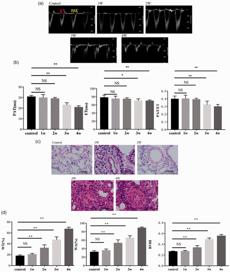
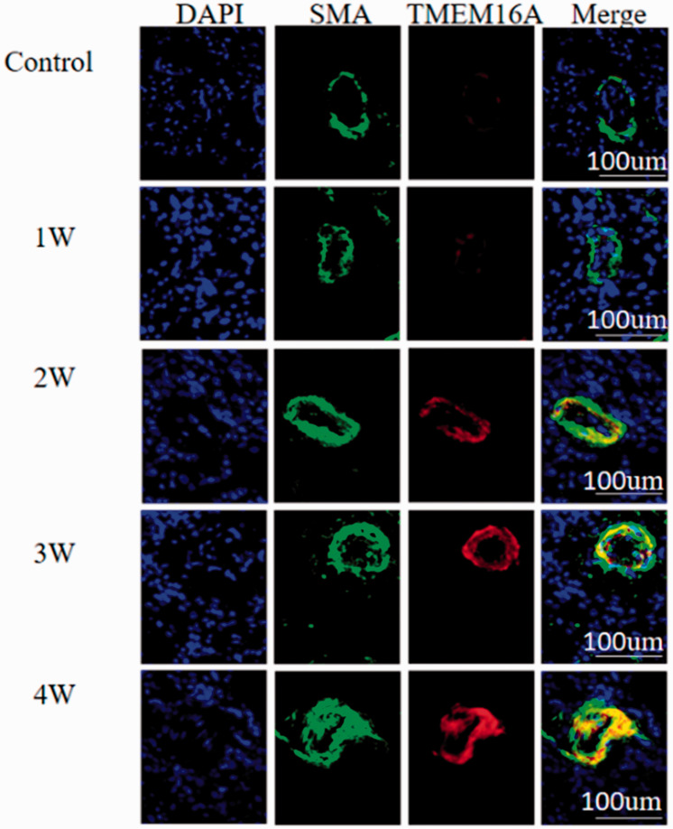
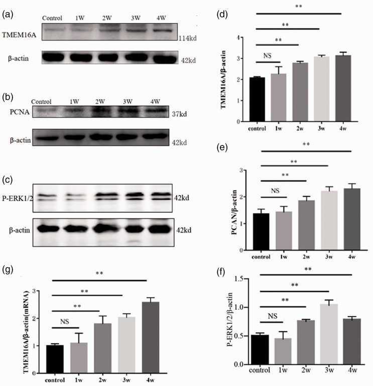
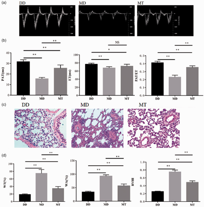
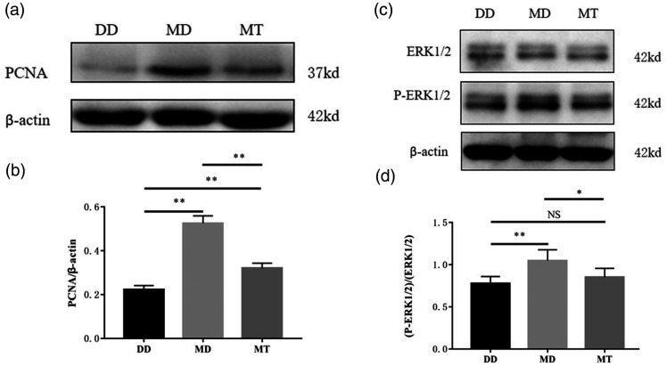
Transmembrane protein 16A was involved in the development of the monocrotaline-induced pulmonary arterial hypertension model through ERK1/2 activation, and it was considered as potential target for pulmonary arterial hypertension treatment. A pulmonary arterial hypertension rat model was established by intraperitoneal administration of monocrotaline. Noninvasive pulsed-wave Doppler and histological analysis was performed, and it revealed proliferation and remodeling of pulmonary arterioles and right ventricle hypertrophy. In addition, transmembrane protein 16A, proliferating cell nuclear antigen-a proliferate marker, P-ERK1/2 increased following monocrotaline treatment. Expression of transmembrane protein 16A in the pulmonary arteries was co-localized with a specific marker of vascular smooth muscle α-actin. Then, a specific inhibitor of transmembrane protein 16A-T16A-A01 was administered to pulmonary arterial hypertension rats. It was found to alleviate the remodeling of pulmonary arterioles and right ventricle hypertrophy significantly, and decrease the upregulation of proliferating cell nuclear antigen in monocrotaline-induced pulmonary arteries. In addition, T16A-A01 could inhibit the activation of ERK1/2 in pulmonary arterial hypertension model. Transmembrane protein 16A mediated the proliferation and remodeling of pulmonary arterioles in the monocrotaline-induced pulmonary arterial hypertension model. ERK1/2 pathway is one of downstream factors. Long-term use of T16A-A01 in vivo could alleviate remodeling and pressure in pulmonary arterial hypertension.
跨膜蛋白16A通过激活ERK1/2参与了野百合碱诱导的肺动脉高压模型的发展,并且它被认为是肺动脉高压治疗的潜在靶点。通过腹腔注射野百合碱建立了肺动脉高压大鼠模型。进行了无创脉冲波多普勒和组织学分析,结果显示肺小动脉增殖和重塑以及右心室肥大。此外,野百合碱处理后跨膜蛋白16A、增殖细胞核抗原(一种增殖标志物)、磷酸化ERK1/2增加。肺血管中跨膜蛋白16A的表达与血管平滑肌α-肌动蛋白的特异性标志物共定位。然后,将跨膜蛋白16A的特异性抑制剂T16A-A01给予肺动脉高压大鼠。发现它能显著减轻肺小动脉重塑和右心室肥大,并降低野百合碱诱导的肺动脉中增殖细胞核抗原的上调。此外,T16A-A01可以抑制肺动脉高压模型中ERK1/2的激活。在野百合碱诱导的肺动脉高压模型中,跨膜蛋白16A介导了肺小动脉的增殖和重塑。ERK1/2途径是下游因素之一。在体内长期使用T16A-A01可以减轻肺动脉高压中的重塑和压力。