• 文献检索
  • 文档翻译
  • 深度研究
  • 学术资讯
  • Suppr Zotero 插件Zotero 插件
  • 邀请有礼
  • 套餐&价格
  • 历史记录
应用&插件
Suppr Zotero 插件Zotero 插件浏览器插件Mac 客户端Windows 客户端微信小程序
定价
高级版会员购买积分包购买API积分包
服务
文献检索文档翻译深度研究API 文档MCP 服务
关于我们
关于 Suppr公司介绍联系我们用户协议隐私条款
关注我们

Suppr 超能文献

核心技术专利:CN118964589B侵权必究
粤ICP备2023148730 号-1Suppr @ 2026

文献检索

告别复杂PubMed语法,用中文像聊天一样搜索,搜遍4000万医学文献。AI智能推荐,让科研检索更轻松。

立即免费搜索

文件翻译

保留排版,准确专业,支持PDF/Word/PPT等文件格式,支持 12+语言互译。

免费翻译文档

深度研究

AI帮你快速写综述,25分钟生成高质量综述,智能提取关键信息,辅助科研写作。

立即免费体验

圆锥角膜交联手术后无菌性角膜浸润患者的回顾性分析

Retrospective Analysis of Sterile Corneal Infiltrates in Patients with Keratoconus after Cross-Linking Procedure.

作者信息

Krok Magdalena, Wróblewska-Czajka Ewa, Kokot Joanna, Micińska Anna, Wylęgała Edward, Dobrowolski Dariusz

机构信息

Chair and Clinical Department of Ophthalmology, Faculty of Medical Sciences in Zabrze, Medical University of Silesia, Panewnicka 65 Street, 40-760 Katowice, Poland.

Ophthalmology of Department, District Railway Hospital, Panewnicka 65 Street, 40-760 Katowice, Poland.

出版信息

J Clin Med. 2022 Jan 25;11(3):585. doi: 10.3390/jcm11030585.

DOI:10.3390/jcm11030585
PMID:35160037
原文链接:https://pmc.ncbi.nlm.nih.gov/articles/PMC8836725/
Abstract

BACKGROUND

This paper's objective is to analyze patients with keratoconus who developed sterile infiltrate after corneal collagen cross-linking (CXL), and to evaluate possible risk factors for their occurrence.

METHODS

543 medical histories of patients after cross-linking (Epi-off, Epi-on) procedure performed according to the Dresden protocol were analyzed retrospectively.

RESULTS

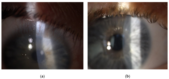
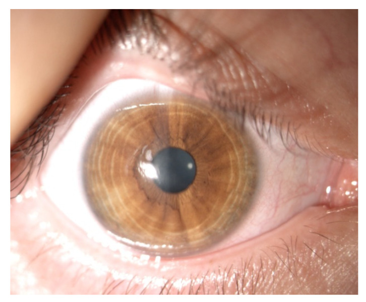
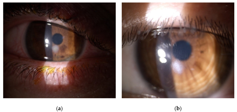
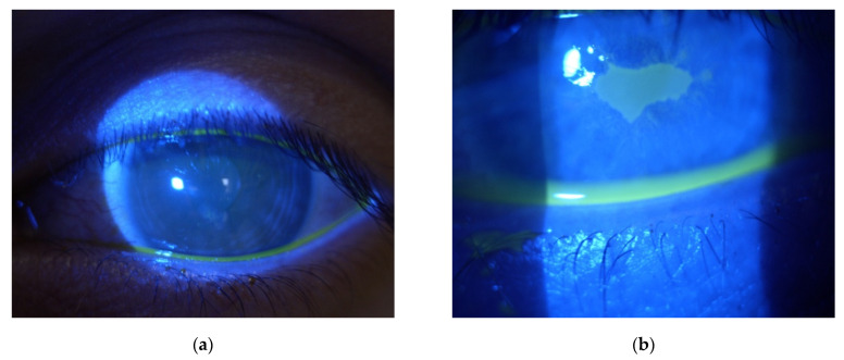
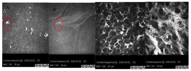
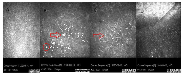
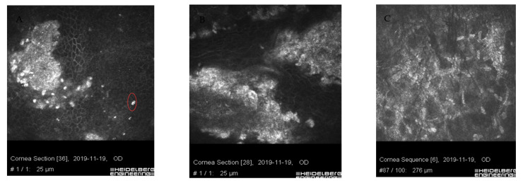
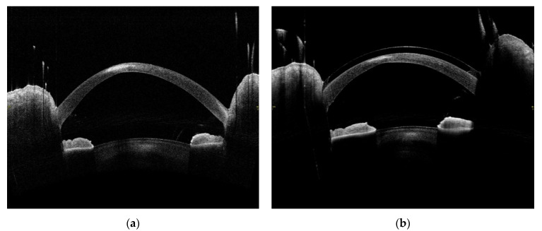
Sterile corneal infiltrates occurred in four men (0.7%) in the age range (16-28) years, the average age being 20.3. The average time from procedure to onset of symptoms was 3.5 days (2-5 days). Inflammatory infiltration resolved in all patients, leaving scars on corneal stroma in two patients. Corneal healing time ranged from 4-12 weeks. In vivo confocal microscopy (IVCM), round inflammatory cells, and Langerhans cells in the epithelium and Bowman's layer were observed at the site of infiltration. The Optical coherence tomography (OCT) shows hyperreflective lesions of various sizes which decreased over time. The corneal topographic parameters and Best-corrected visual acuity (BCVA) improved after the CXL procedure in all of the described cases.

CONCLUSIONS

Most likely, damage to the epithelium and the phototoxic effect of the procedure is of significant importance in the formation of sterile corneal infiltrates. Appropriate classification and selection of CXL procedures in combination with protective measures in people at risk may have an overwhelming impact on the incidence of this complication.

摘要

背景

本文旨在分析圆锥角膜患者在角膜胶原交联(CXL)后发生无菌性浸润的情况,并评估其发生的可能危险因素。

方法

回顾性分析了543例按照德累斯顿方案进行交联手术(上皮去除、上皮保留)后的患者病历。

结果

4名男性(0.7%)出现无菌性角膜浸润,年龄在16 - 28岁之间,平均年龄为20.3岁。从手术到症状出现的平均时间为3.5天(2 - 5天)。所有患者的炎症浸润均消退,2例患者角膜基质留下瘢痕。角膜愈合时间为4 - 12周。在浸润部位通过活体共聚焦显微镜(IVCM)观察到上皮和Bowman层中有圆形炎症细胞和朗格汉斯细胞。光学相干断层扫描(OCT)显示大小各异的高反射性病变,且随时间减少。在所有上述病例中,CXL手术后角膜地形图参数和最佳矫正视力(BCVA)均有所改善。

结论

上皮损伤和手术的光毒性作用很可能在无菌性角膜浸润的形成中起重要作用。对CXL手术进行适当分类和选择,并对高危人群采取保护措施,可能会对该并发症的发生率产生重大影响。

https://cdn.ncbi.nlm.nih.gov/pmc/blobs/dca2/8836725/158d9840b5f0/jcm-11-00585-g008.jpg
https://cdn.ncbi.nlm.nih.gov/pmc/blobs/dca2/8836725/9754bc6ecb77/jcm-11-00585-g001.jpg
https://cdn.ncbi.nlm.nih.gov/pmc/blobs/dca2/8836725/d243b6ed7da0/jcm-11-00585-g002.jpg
https://cdn.ncbi.nlm.nih.gov/pmc/blobs/dca2/8836725/12bda2807471/jcm-11-00585-g003.jpg
https://cdn.ncbi.nlm.nih.gov/pmc/blobs/dca2/8836725/f55e84c5d2ca/jcm-11-00585-g004.jpg
https://cdn.ncbi.nlm.nih.gov/pmc/blobs/dca2/8836725/3a178a4a359a/jcm-11-00585-g005.jpg
https://cdn.ncbi.nlm.nih.gov/pmc/blobs/dca2/8836725/8da5f3782b56/jcm-11-00585-g006.jpg
https://cdn.ncbi.nlm.nih.gov/pmc/blobs/dca2/8836725/9c70b3aec6ef/jcm-11-00585-g007.jpg
https://cdn.ncbi.nlm.nih.gov/pmc/blobs/dca2/8836725/158d9840b5f0/jcm-11-00585-g008.jpg

相似文献

1
Retrospective Analysis of Sterile Corneal Infiltrates in Patients with Keratoconus after Cross-Linking Procedure.圆锥角膜交联手术后无菌性角膜浸润患者的回顾性分析
J Clin Med. 2022 Jan 25;11(3):585. doi: 10.3390/jcm11030585.
2
Sterile corneal infiltrates after corneal collagen cross-linking: evaluation of risk factors.角膜胶原交联术后的无菌性角膜浸润:危险因素评估
Acta Ophthalmol. 2017 Mar;95(2):199-204. doi: 10.1111/aos.13218. Epub 2016 Oct 24.
3
Collagen cross-linking using riboflavin and ultraviolet-a for corneal thinning disorders: an evidence-based analysis.使用核黄素和紫外线A进行角膜交联治疗角膜变薄疾病:一项循证分析
Ont Health Technol Assess Ser. 2011;11(5):1-89. Epub 2011 Nov 1.
4
Accelerated epithelium-on or accelerated epithelium-off corneal collagen cross-linking: Contralateral comparison study.加速上皮在位或加速上皮脱失的角膜胶原交联:对侧比较研究。
Taiwan J Ophthalmol. 2020 Mar 4;10(1):37-44. doi: 10.4103/tjo.tjo_11_19. eCollection 2020 Jan-Mar.
5
Transepithelial versus epithelium-off corneal cross-linking for the treatment of progressive keratoconus: a randomized controlled trial.经上皮与上皮下角膜交联治疗进行性圆锥角膜的随机对照试验
Am J Ophthalmol. 2015 May;159(5):821-8.e3. doi: 10.1016/j.ajo.2015.02.005. Epub 2015 Feb 19.
6
Iontophoretic collagen cross-linking versus epithelium-off collagen cross-linking for early stage of progressive keratoconus - 3 years follow-up study.离子导入胶原交联与上皮去除胶原交联治疗早期进行性圆锥角膜的3年随访研究
Acta Ophthalmol. 2017 Nov;95(7):e649-e655. doi: 10.1111/aos.13538.
7
Fourier-domain optical coherence tomography imaging in keratoconus: a corneal structural classification.频域光学相干断层成像在圆锥角膜中的应用:角膜结构分类。
Ophthalmology. 2013 Dec;120(12):2403-2412. doi: 10.1016/j.ophtha.2013.05.027. Epub 2013 Aug 9.
8
Epithelial and stromal remodeling after corneal collagen cross-linking evaluated by spectral-domain OCT.应用频域光相干断层扫描评估角膜胶原交联术后上皮和基质重塑。
J Refract Surg. 2014 Feb;30(2):122-7. doi: 10.3928/1081597X-20140120-08.
9
[Transepithelial iontophoresis corneal collagen cross-linking for progressive keratoconus: one year results].[经上皮离子导入角膜胶原交联治疗进展性圆锥角膜:一年结果]
Zhonghua Yan Ke Za Zhi. 2015 Sep;51(9):677-82.
10
Peripheral sterile corneal ring infiltrate after riboflavin-UVA collagen cross-linking in keratoconus.圆锥角膜行核黄素-UVA 胶原交联术后周边无菌性角膜环浸润。
Cornea. 2012 Jun;31(6):702-5. doi: 10.1097/ICO.0b013e318226da53.

引用本文的文献

1
Keratoconus: exploring fundamentals and future perspectives - a comprehensive systematic review.圆锥角膜:探索基础与未来展望——一项全面的系统综述
Ther Adv Ophthalmol. 2024 Mar 20;16:25158414241232258. doi: 10.1177/25158414241232258. eCollection 2024 Jan-Dec.
2
Severe Complications after Corneal Collagen Cross-Linking (CXL).严重并发症后的角膜胶原交联 (CXL)。
Klin Monbl Augenheilkd. 2023 Apr;240(4):369-378. doi: 10.1055/a-2040-4290. Epub 2023 Apr 25.

本文引用的文献

1
Visual outcomes of sterile corneal infiltrates after photorefractive keratectomy.光折射性角膜切削术后无菌性角膜浸润的视力结果。
Indian J Ophthalmol. 2020 Dec;68(12):2956-2959. doi: 10.4103/ijo.IJO_1300_20.
2
Keratitis following laser refractive surgery: Clinical spectrum, prevention and management.激光屈光手术后的角膜炎:临床谱、预防与管理
Indian J Ophthalmol. 2020 Dec;68(12):2813-2818. doi: 10.4103/ijo.IJO_2479_20.
3
Complications of accelerated corneal collagen cross-linking: review of 2025 eyes.加速角膜胶原交联术的并发症:2025 只眼的回顾。
Int Ophthalmol. 2020 Dec;40(12):3269-3277. doi: 10.1007/s10792-020-01512-3. Epub 2020 Jul 26.
4
Profile of infectious and sterile keratitis after accelerated corneal collagen cross-linking for keratoconus.圆锥角膜加速角膜胶原交联术后感染性和非感染性角膜炎的概况
Oman J Ophthalmol. 2020 Feb 17;13(1):18-23. doi: 10.4103/ojo.OJO_115_2018. eCollection 2020 Jan-Apr.
5
Sterile keratitis following standard corneal collagen crosslinking: A case series and literature review.标准角膜胶原交联术后无菌性角膜炎:病例系列及文献综述
J Fr Ophtalmol. 2019 Jun;42(6):603-611. doi: 10.1016/j.jfo.2018.11.015. Epub 2019 Apr 5.
6
Sterile keratitis after uneventful corneal collagen cross-linking in a patient with Axenfeld-Rieger syndrome.一名患有Axenfeld-Rieger综合征的患者在角膜胶原交联术后发生无菌性角膜炎。
Int Ophthalmol. 2019 May;39(5):1169-1173. doi: 10.1007/s10792-018-0907-1. Epub 2018 Mar 28.
7
Sterile corneal infiltrates after corneal collagen cross-linking: evaluation of risk factors.角膜胶原交联术后的无菌性角膜浸润:危险因素评估
Acta Ophthalmol. 2017 Mar;95(2):199-204. doi: 10.1111/aos.13218. Epub 2016 Oct 24.
8
Progress in corneal wound healing.角膜伤口愈合的进展。
Prog Retin Eye Res. 2015 Nov;49:17-45. doi: 10.1016/j.preteyeres.2015.07.002. Epub 2015 Jul 18.
9
In Vivo Confocal Microscopy after Corneal Collagen Crosslinking.角膜胶原交联术后的体内共聚焦显微镜检查
Ocul Surf. 2015 Oct;13(4):298-314. doi: 10.1016/j.jtos.2015.04.007. Epub 2015 Jul 2.
10
Sterile Keratitis following Collagen Crosslinking.胶原交联术后的无菌性角膜炎
J Ophthalmic Vis Res. 2014 Oct-Dec;9(4):510-3. doi: 10.4103/2008-322X.150832.