Department of Molecular and Cellular Biology, Faculty of Pharmacy, Wroclaw Medical University, Borowska 211A St., 50-556 Wroclaw, Poland.
Department of Engineering and Technology of Chemical Processes, Faculty of Chemistry, Wroclaw University of Technology, Wybrzeze Wyspianskiego 27, 50-370 Wroclaw, Poland.
Int J Mol Sci. 2022 Jan 26;23(3):1377. doi: 10.3390/ijms23031377.
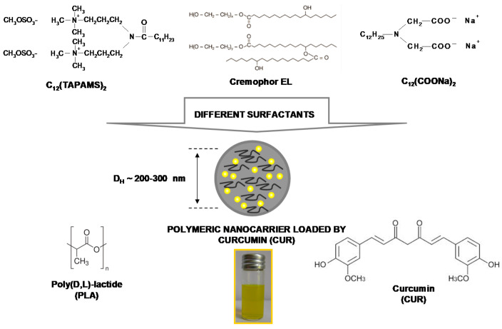
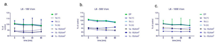
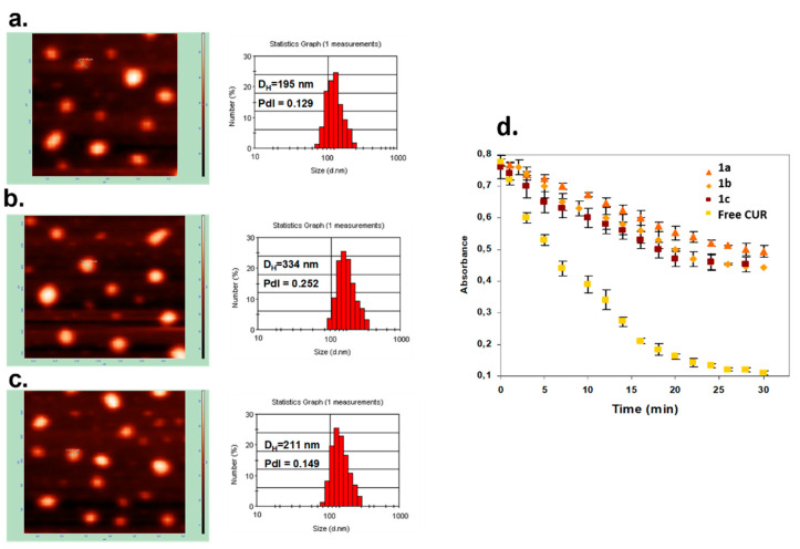
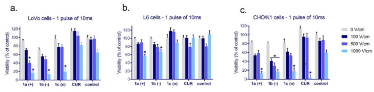
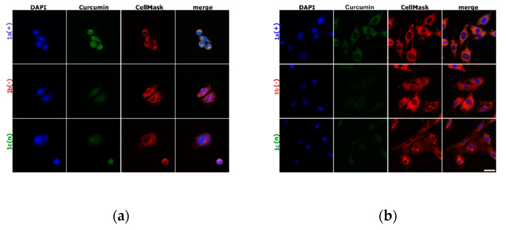
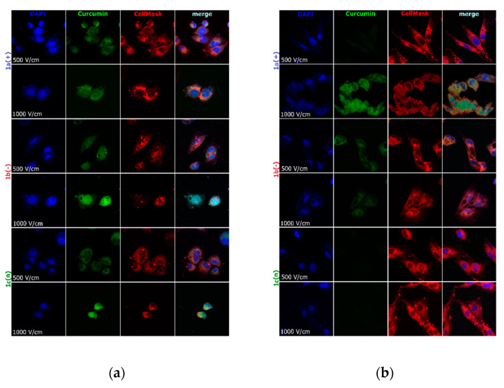
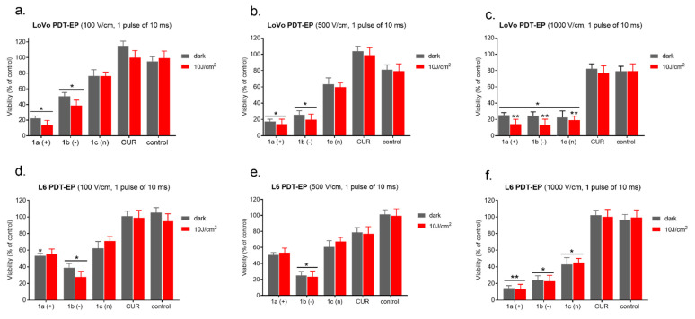
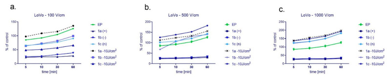
(1) Background: The size and surface charge are the most significant parameters of nanocarriers that determine their efficiency and potential application. The poor cell uptake of encapsulated drugs is the main limitation in anticancer treatment. The well-defined properties of nanocarriers will enable to target specific tissue and deliver an active cargo. (2) Methods: In the current study, poly(D,L -lactide) (PLA) nanocarriers loaded with curcumin (CUR) and differing surface charge were evaluated for transport efficacy in combination with electroporation (EP) in dependence on the type of cells. The obtained CUR-loaded nanoparticles with diameters ranging from 195 to 334 nm (derived from dynamic light scattering (DLS)) were characterized by atomic force microscopy (AFM) (morphology and shape) and Doppler electrophoresis (-potential) as well as UV-vis spectroscopy (CUR encapsulation efficiency (about 90%) and photobleaching rate). The drug delivery properties of the obtained PLA nanocarriers enhanced by electroporation were assessed in human colon cancer cells (LoVo), excitable normal rat muscle cells (L6), and free of voltage-gated ion channels cells (CHO-K1). CLSM studies, viability, and ROS release were performed to determine the biological effects of nanocarriers. (3) Results: The highest photodynamic activity indicated anionic nanocarriers (1a) stabilized by C(COONa) surfactant. Nanocarriers were cytotoxic for LoVo cells and less cytotoxic for normal cells. ROS release increased in cancer cells with the increasing electric field intensity, irradiation, and time after EP. Muscle L6 cells were less sensitive to electric pulses. (4) Conclusions: EP stimulation for CUR-PLA nanocarriers transport was considered to improve the regulated and more effective delivery of nanosystems differing in surface charge.
(1) 背景:纳米载体的大小和表面电荷是决定其效率和潜在应用的最重要参数。包裹药物的细胞摄取效率低下是癌症治疗的主要限制。纳米载体的明确特性将能够靶向特定组织并输送有效载荷。(2) 方法:在当前的研究中,载有姜黄素 (CUR) 且表面电荷不同的聚(D,L-乳酸) (PLA) 纳米载体与电穿孔 (EP) 联合用于评估其在不同类型细胞中的输送效果。通过原子力显微镜 (AFM) (形态和形状) 和多普勒电泳 (-电位) 以及紫外可见光谱 (CUR 包封效率 (约 90%) 和光漂白率) 对获得的直径在 195 至 334nm 之间的 CUR 负载纳米颗粒 (源自动态光散射 (DLS)) 进行了表征。通过电穿孔增强的 PLA 纳米载体的药物传递特性在人结肠癌细胞 (LoVo)、可兴奋的正常大鼠肌肉细胞 (L6) 和无电压门控离子通道细胞 (CHO-K1) 中进行了评估。通过共聚焦激光扫描显微镜 (CLSM) 研究、细胞活力和 ROS 释放来确定纳米载体的生物学效应。(3) 结果:带负电荷的纳米载体 (1a) 显示出最高的光动力活性,由 C(COONa) 表面活性剂稳定。纳米载体对 LoVo 细胞具有细胞毒性,对正常细胞的细胞毒性较小。随着电场强度、辐照和 EP 后时间的增加,ROS 在癌细胞中的释放增加。肌肉 L6 细胞对电脉冲的敏感性较低。(4) 结论:考虑对 CUR-PLA 纳米载体进行 EP 刺激以改善表面电荷不同的纳米系统的调节和更有效的输送。