State Key Laboratory of Quality Research in Chinese Medicine, Institute of Chinese Medical Sciences, University of Macau, Macao SAR 999078, China.
Department of Pharmaceutical Sciences, Faculty of Health Sciences, University of Macau, Macao SAR 999078, China.
Int J Mol Sci. 2022 Feb 7;23(3):1878. doi: 10.3390/ijms23031878.
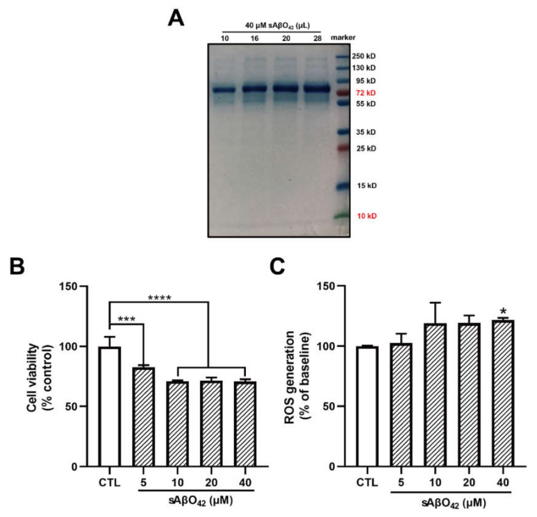
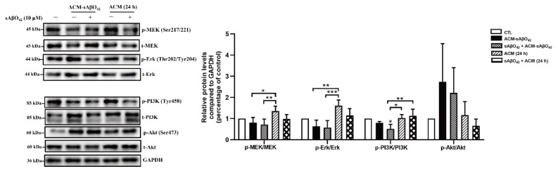
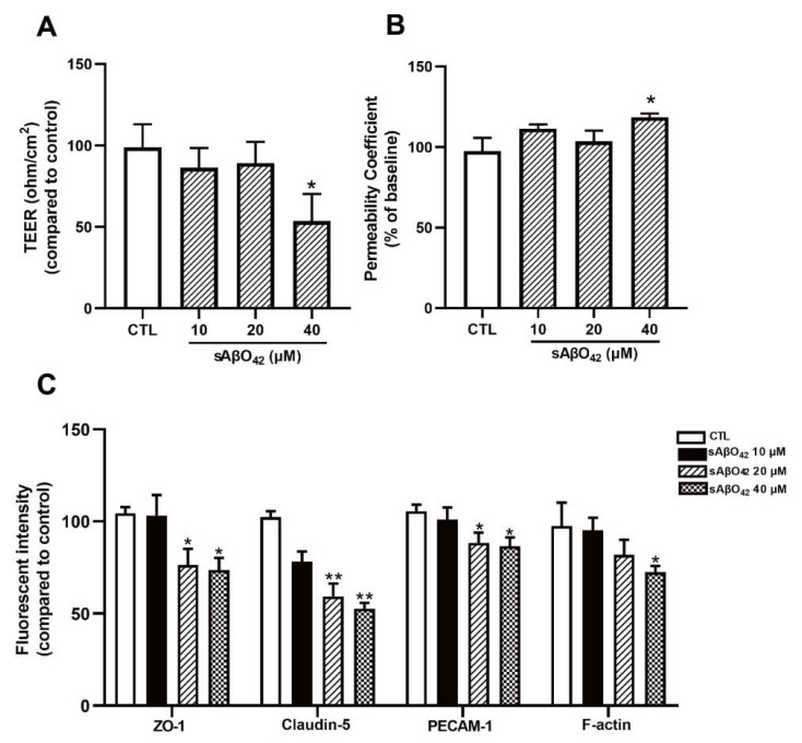
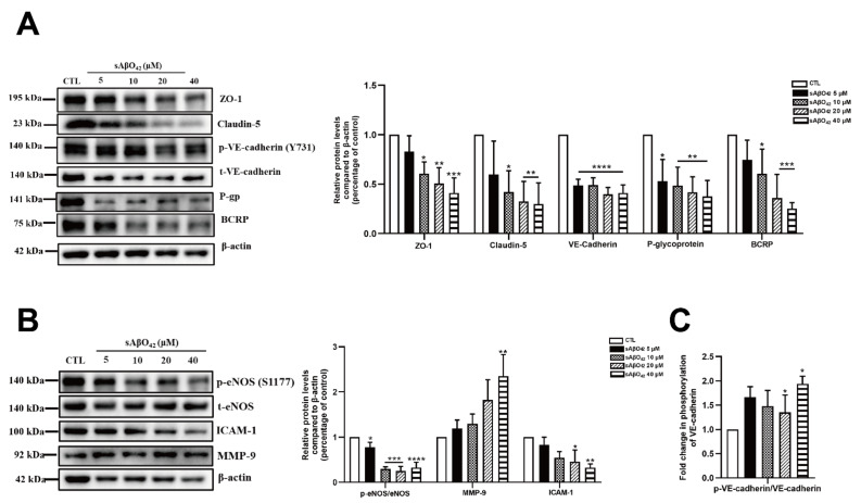
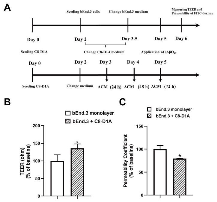
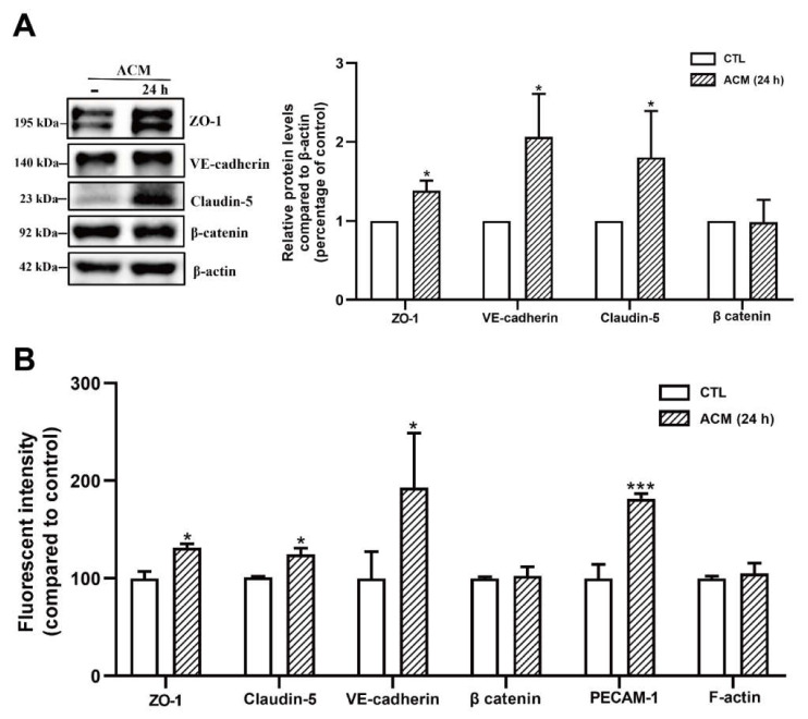
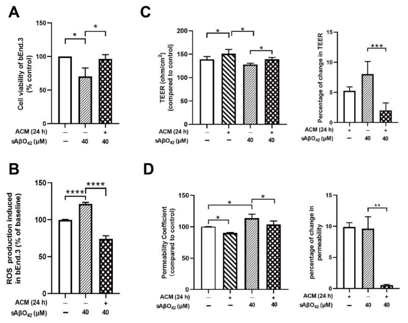
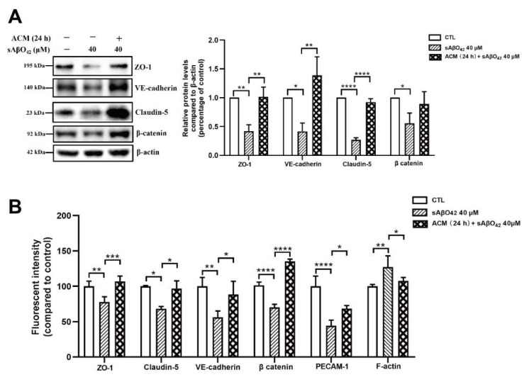
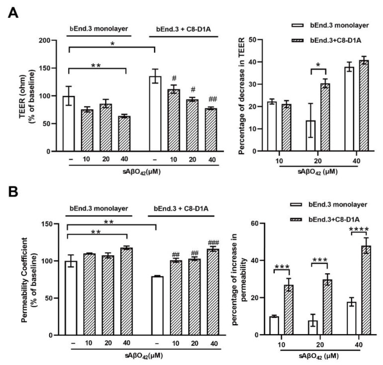
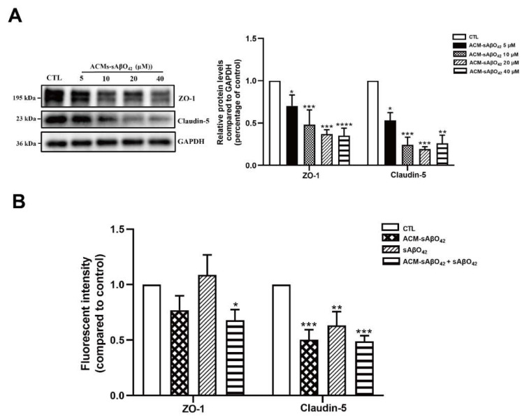
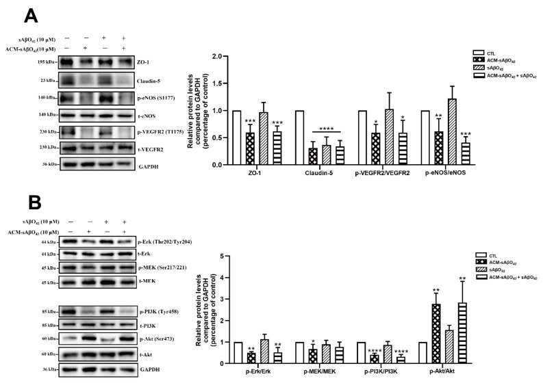
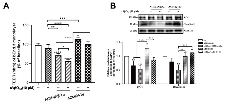
Transgenic mouse models of Alzheimer's disease (AD) overexpress mutations of the human amyloid protein precursor () and presenilin-1 () genes, which are known causes of amyloid pathology in familial AD. However, animal models for studying AD in the context of aging and age-related co-morbidities, such as blood-brain barrier (BBB) disruptions, are lacking. More recently, aged and progeroid mouse models have been proposed as alternatives to study aging-related AD, but the toxicity of murine amyloid-beta protein (Aβ) is not well defined. In this study, we aimed to study the potential toxicity of murine Aβ on brain endothelial cells and astrocytes, which are important components of the BBB, using mouse brain endothelial cells (bEnd.3) and astrocytes (C8-D1A). Murine-soluble Aβ (1-42) oligomers (sAβO42) (10 µM) induced negligible injuries in an endothelial monolayer but induced significant barrier disruptions in a bEnd.3 and C8-D1A co-culture. Similar results of endothelial perturbation were observed in a bEnd.3 monolayer treated with astrocyte-conditioned medium (ACM) generated by astrocytes exposed to sAβO42 (ACM-sAβO42), while additional exogenous sAβO42 did not cause further damage. Western blot analysis showed that ACM-sAβO42 altered the basal activities of vascular endothelial growth factor receptor 2 (VEGFR2), eNOS, and the signaling of the MEK/ERK and Akt pathways in bEnd.3. Our results showed that murine sAβO42 was moderately toxic to an endothelial and astrocyte co-culture. These damaging effects on the endothelial barrier were induced by deleterious soluble factors released from astrocytes, which disrupted endothelial VEGFR2 signaling and perturbed cell survival and barrier stabilization.
阿尔茨海默病(AD)的转基因小鼠模型过度表达人类淀粉样蛋白前体()和早老素-1()基因的突变,这些突变已知是家族性 AD 中淀粉样蛋白病理学的原因。然而,缺乏用于研究与衰老和与年龄相关的合并症(如血脑屏障(BBB)破坏)相关的 AD 的动物模型。最近,已经提出了老龄和早衰小鼠模型作为研究与衰老相关的 AD 的替代方法,但鼠淀粉样β蛋白(Aβ)的毒性尚未得到很好的定义。在这项研究中,我们旨在使用小鼠脑内皮细胞(bEnd.3)和星形胶质细胞(C8-D1A)研究脑内皮细胞和星形胶质细胞中鼠 Aβ 的潜在毒性,脑内皮细胞和星形胶质细胞是 BBB 的重要组成部分。鼠可溶性 Aβ(1-42)寡聚物(sAβO42)(10 µM)在内皮单层中引起的损伤可以忽略不计,但在 bEnd.3 和 C8-D1A 共培养物中引起明显的屏障破坏。在用暴露于 sAβO42 的星形胶质细胞产生的星形胶质细胞条件培养基(ACM-sAβO42)处理的 bEnd.3 单层中观察到类似的内皮扰动结果,而额外的外源性 sAβO42 不会造成进一步的损伤。Western blot 分析显示,ACM-sAβO42 改变了 bEnd.3 中血管内皮生长因子受体 2(VEGFR2)、eNOS 的基础活性以及 MEK/ERK 和 Akt 通路的信号转导。我们的结果表明,鼠 sAβO42 对内皮和星形胶质细胞共培养物具有中度毒性。这些对内皮屏障的破坏作用是由星形胶质细胞释放的有害可溶性因子引起的,这些因子破坏了内皮 VEGFR2 信号转导并扰乱了细胞存活和屏障稳定性。