Nara Institute of Science and Technology, Graduate School of Science and Technology, Division of Mat, Japan.
Kumamoto University, Graduate School of Pharmaceutical Sciences, Department of Chemico-Pharmacologic, Japan.
J Biomed Opt. 2022 Feb;27(2). doi: 10.1117/1.JBO.27.2.026501.
Intrinsic optical signals (IOS) generated in the cortical tissue as a result of various interacting metabolic processes are used extensively to elucidate the underlying mechanisms that govern neurovascular coupling. However, current IOS measurements still often rely on bulky, tabletop imaging systems, and there remains a dearth of studies in freely moving subjects. Lightweight, miniature head-mounted imaging devices provide unique opportunities for investigating cortical dynamics in small animals under a variety of naturalistic behavioral settings.
The aim of this work was to monitor IOS in the somatosensory cortex of wild-type mice by developing a lightweight, biocompatible imaging device that readily lends itself to animal experiments in freely moving conditions.
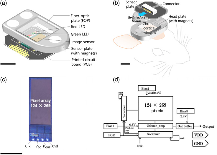
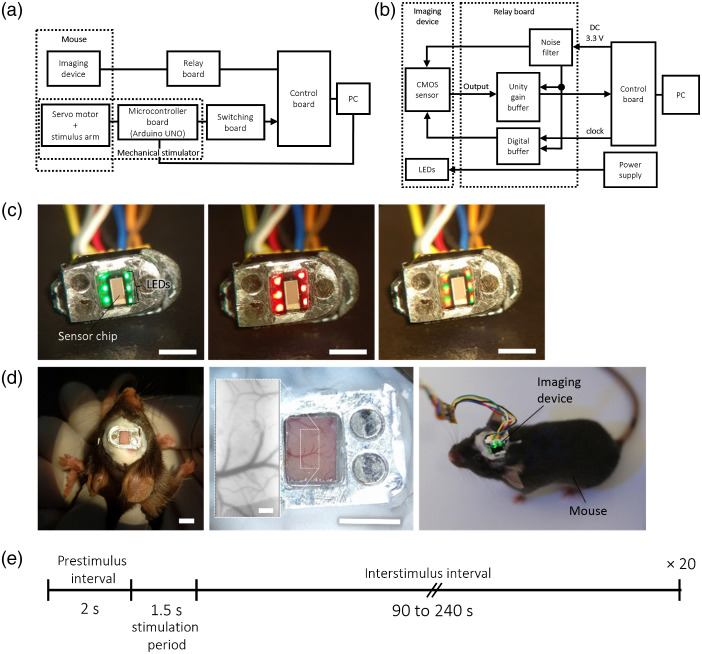
Herein we describe a method for realizing long-term IOS imaging in mice using a 0.54-g, compact, CMOS-based, head-mounted imager. The two-part module, consisting of a tethered sensor plate and a base plate, allows facile assembly prior to imaging sessions and disassembly when the sensor is not in use. LEDs integrated into the device were chosen to illuminate the cortical mantle at two different wavelengths in the visible regime (λcenter: 535 and 625 nm) for monitoring volume- and oxygenation state-dependent changes in the IOS, respectively. To test whether the system can detect robust cortical responses, we recorded sensory-evoked IOS from mechanical stimulation of the hindlimbs (HL) of anesthetized mice in both acute and long-term implantation conditions.
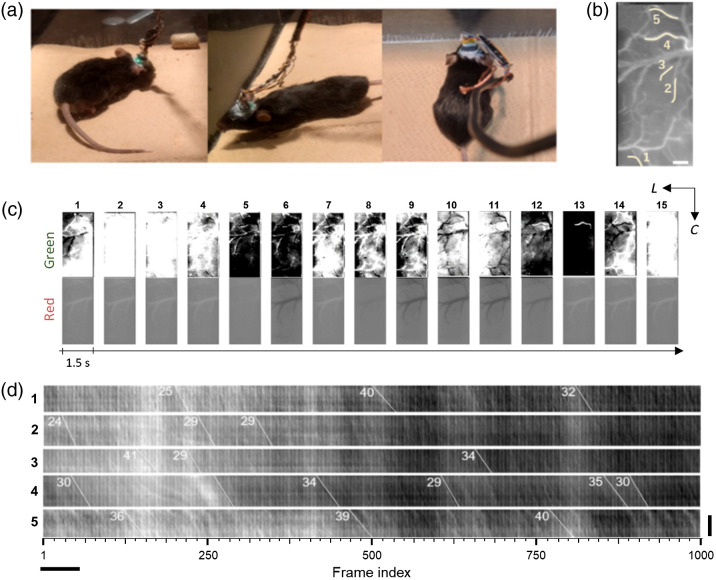
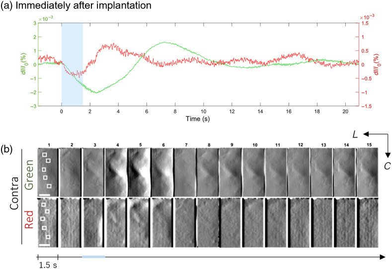
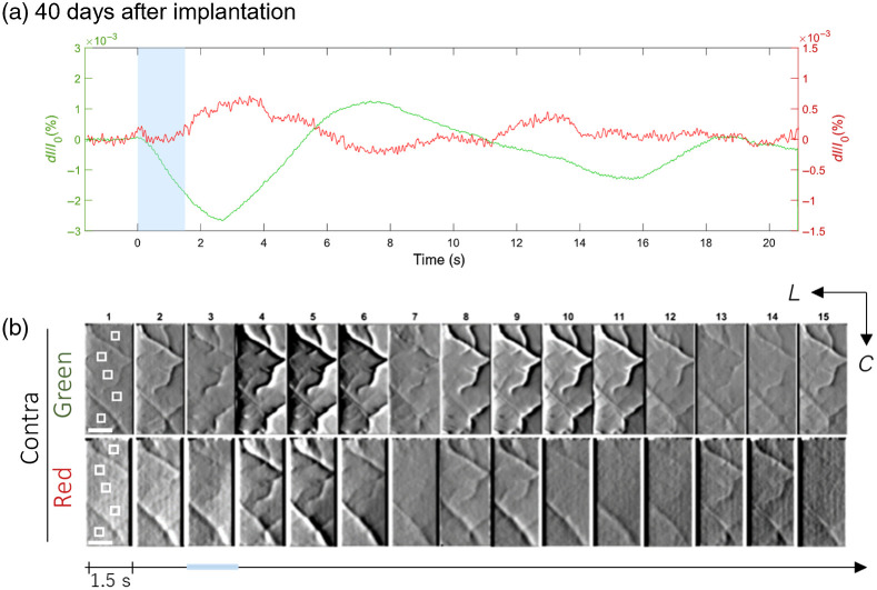
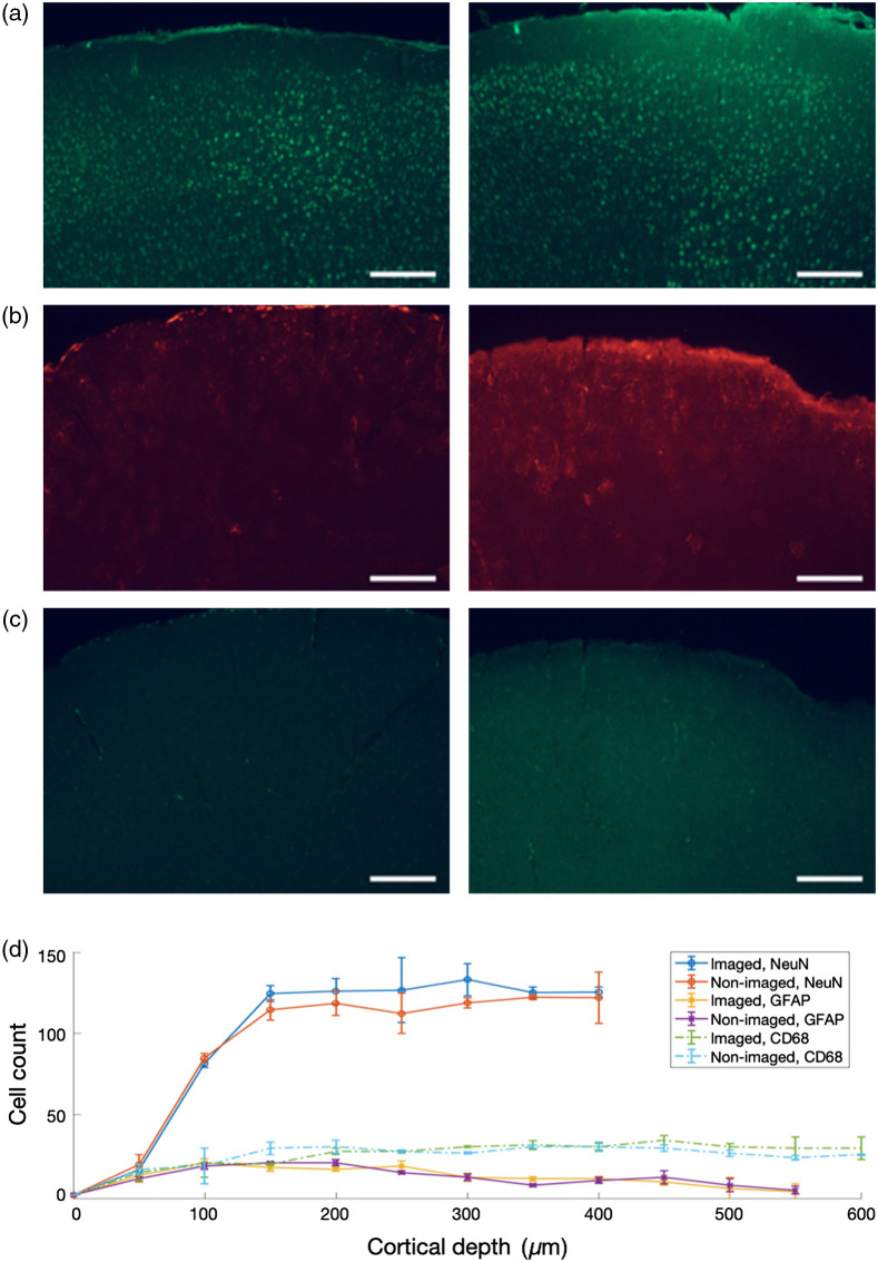
Cortical IOS recordings in the primary somatosensory cortex hindlimb receptive field (S1HL) of anesthetized mice under green and red LED illumination revealed robust, multiphasic profiles that were time-locked to the mechanical stimulation of the contralateral plantar hindpaw. Similar intrinsic signal profiles observed in S1HL at 40 days postimplantation demonstrated the viability of the approach for long-term imaging. Immunohistochemical analysis showed that the brain tissue did not exhibit appreciable immune response due to the device implantation and operation. A proof-of-principle imaging session in a freely behaving mouse showed minimal locomotor impediment for the animal and also enabled estimation of blood flow speed.
We demonstrate the utility of a miniature cortical imaging device for monitoring IOS and related hemodynamic processes in both anesthetized and freely moving mice, cueing potential for applications to some neuroscientific studies of sensation and naturalistic behavior.
作为各种相互作用的代谢过程的结果,在皮质组织中产生的固有光学信号 (IOS) 被广泛用于阐明支配神经血管耦合的基本机制。然而,当前的 IOS 测量仍然经常依赖于庞大的台式成像系统,并且在自由移动的对象中仍然缺乏研究。轻便、微型的头戴式成像设备为在各种自然行为环境下研究小动物的皮质动力学提供了独特的机会。
本工作旨在通过开发一种轻便、生物相容的成像设备来监测野生型小鼠感觉皮层中的 IOS,该设备易于在自由活动条件下进行动物实验。
本文描述了一种使用 0.54 克、紧凑的基于 CMOS 的头戴式成像器在小鼠中实现长期 IOS 成像的方法。该两部分模块由一个带有传感器的模块和一个带有传感器的底座组成,在成像前可以方便地组装,在不使用传感器时可以拆卸。集成到设备中的 LED 选择在可见光范围内的两个不同波长(λcenter:535 和 625nm)来分别监测 IOS 的体积和氧合状态依赖性变化。为了测试该系统是否能够检测到强大的皮质反应,我们记录了麻醉小鼠后肢机械刺激(HL)引起的感觉诱发 IOS,同时在急性和长期植入条件下进行了记录。
在绿色和红色 LED 照明下,麻醉小鼠初级体感皮层后肢感受野(S1HL)的皮质 IOS 记录显示了与对侧足底后足机械刺激时间锁定的强大、多相的图谱。在植入后 40 天,在 S1HL 中观察到的类似内在信号图谱表明了该方法进行长期成像的可行性。免疫组织化学分析表明,由于设备植入和操作,脑组织没有表现出明显的免疫反应。在自由活动的小鼠中进行的原理验证成像实验表明,该动物的运动受到的阻碍最小,同时也能够估计血流速度。
我们展示了一种微型皮质成像设备在麻醉和自由活动的小鼠中监测 IOS 和相关血液动力学过程的实用性,为感觉和自然行为的一些神经科学研究应用提供了潜力。