Scheppach Wolfgang, Steger Ulrich, Küsters Wolfgang, Wild Vanessa
Medizinische Klinik, Schwerpunkt Gastroenterologie & Rheumatologie, Klinikum Würzburg Mitte, Standort Juliusspital, Salvatorstr. 7, 97074, Würzburg, Deutschland.
Klinik für Chirurgie, Allgemein- und Viszeralchirurgie, Klinikum Würzburg Mitte, Standort Juliusspital, Würzburg, Deutschland.
Internist (Berl). 2022 May;63(5):551-555. doi: 10.1007/s00108-022-01278-z. Epub 2022 Feb 16.
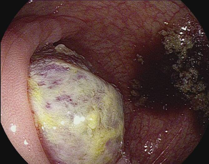
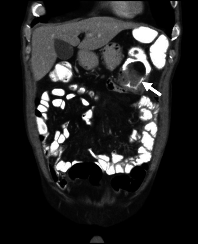
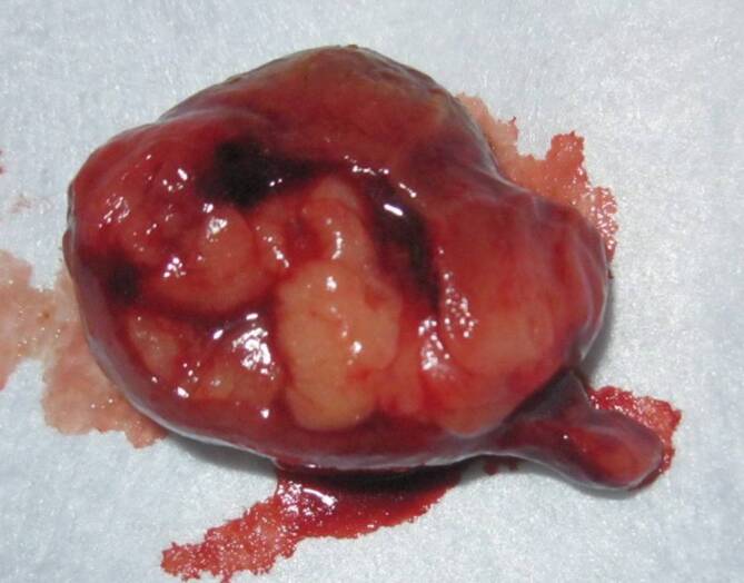
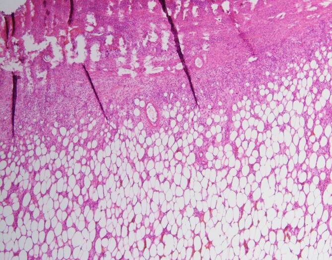
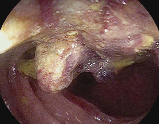
A 61-year-old male patient underwent a colonoscopy for cramp-like upper abdominal pain of 3 weeks duration. An endoscopically irresectable ulcerated mass was seen in the transverse colon. The patient spontaneously excreted in the feces a tumor node measuring 4.1 × 3.5 × 2.8 cm with the histological features of a submucosal lipoma 4 days after the colonoscopy. A benign lipoma confined to the submucosa was operatively confirmed. It is extremely rare for a tumor node to be shed in feces. If the benign nature of the entire lesion is doubtful, standard oncological procedures are advocated.
一名61岁男性患者因持续3周的痉挛样上腹部疼痛接受了结肠镜检查。在横结肠发现一个内镜下无法切除的溃疡性肿块。结肠镜检查4天后,患者粪便中自行排出一个大小为4.1×3.5×2.8 cm的肿瘤结节,具有黏膜下脂肪瘤的组织学特征。手术证实为局限于黏膜下层的良性脂肪瘤。肿瘤结节经粪便排出极为罕见。如果整个病变的良性性质存疑,则提倡采用标准的肿瘤学治疗方法。