Paris-Saclay University, CNRS, Institute of Neuroscience (NeuroPSI), Gif sur Yvette, France.
Center for Interdisciplinary Research in Biology (CIRB), Collège de France, CNRS, INSERM, PSL University, Paris, France.
Biophys J. 2022 Mar 15;121(6):869-885. doi: 10.1016/j.bpj.2022.02.022. Epub 2022 Feb 17.
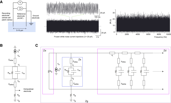
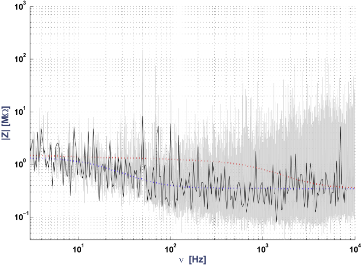
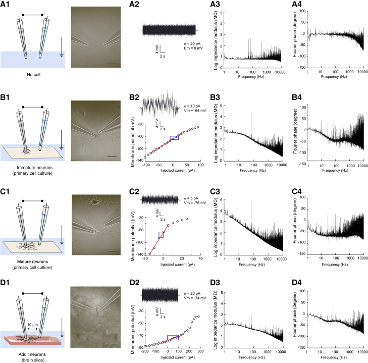
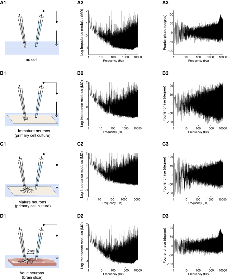
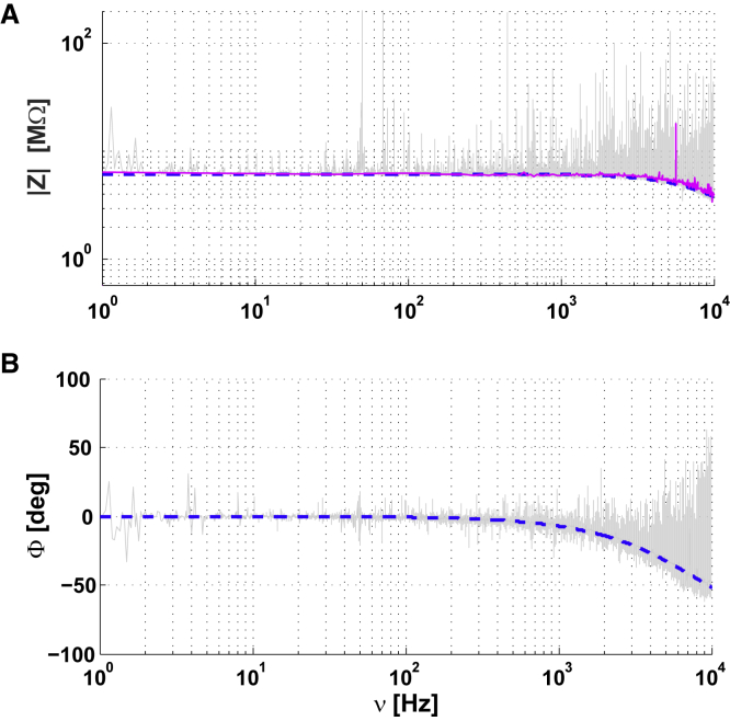
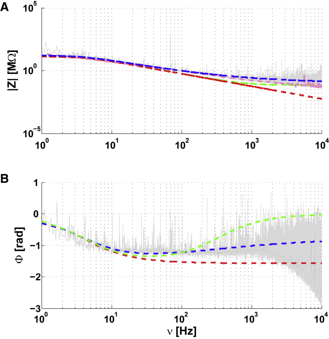
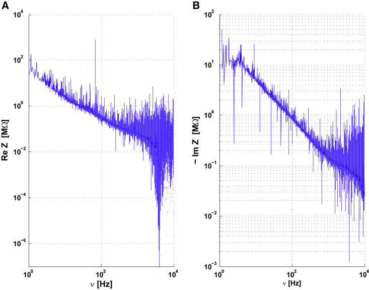
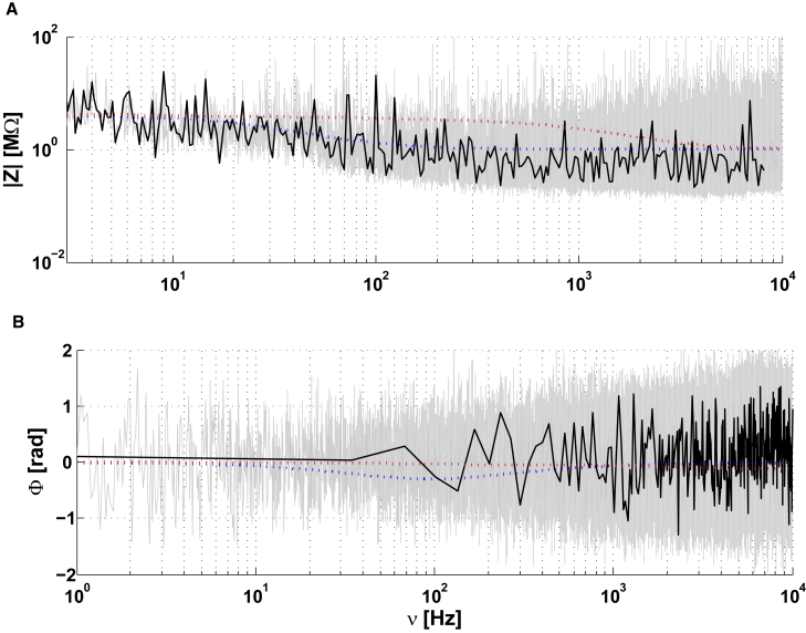
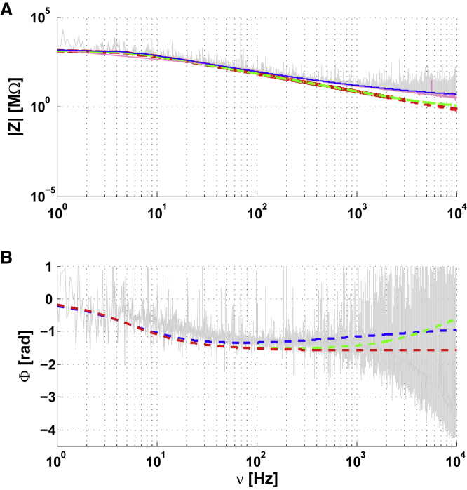
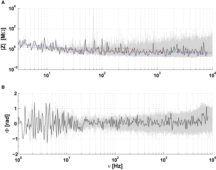
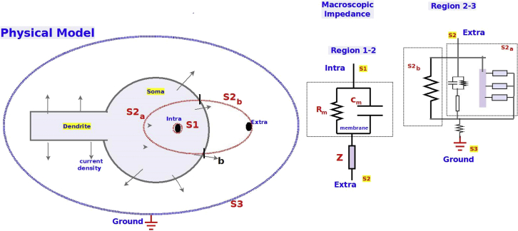
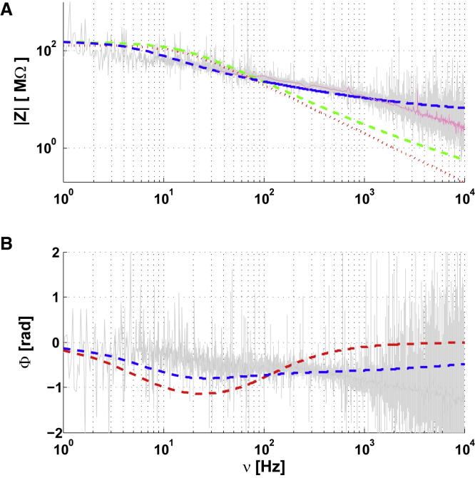
Electric phenomena in brain tissue can be measured using extracellular potentials, such as the local field potential, or the electro-encephalogram. The interpretation of these signals depends on the electric structure and properties of extracellular media, but the measurements of these electric properties are still debated. Some measurements point to a model in which the extracellular medium is purely resistive, and thus parameters such as electric conductivity and permittivity should be independent of frequency. Other measurements point to a pronounced frequency dependence of these parameters, with scaling laws that are consistent with capacitive or diffusive effects. However, these experiments correspond to different preparations, and it is unclear how to correctly compare them. Here, we provide for the first time, impedance measurements (in the 1-10 kHz frequency range) using the same setup in various preparations, from primary cell cultures to acute brain slices, and a comparison with similar measurements performed in artificial cerebrospinal fluid with no biological material. The measurements show that when the current flows across a cell membrane, the frequency dependence of the macroscopic impedance between intracellular and extracellular electrodes is significant, and cannot be captured by a model with resistive media. Fitting a mean-field model to the data shows that this frequency dependence could be explained by the ionic diffusion mainly associated with Debye layers surrounding the membranes. We conclude that neuronal membranes and their ionic environment induce strong deviations to resistivity that should be taken into account to correctly interpret extracellular potentials generated by neurons.
脑组织中的电现象可以通过测量细胞外电势来测量,例如局部场电位或脑电图。这些信号的解释取决于细胞外介质的电结构和特性,但这些电特性的测量仍存在争议。一些测量结果表明,细胞外介质是纯电阻的,因此电导率和介电常数等参数应该与频率无关。其他测量结果表明,这些参数具有明显的频率依赖性,其标度律与电容或扩散效应一致。然而,这些实验对应于不同的制备物,目前尚不清楚如何正确比较它们。在这里,我们首次提供了使用相同设置在各种制备物(从原代细胞培养物到急性脑切片)中进行的阻抗测量(在 1-10 kHz 频率范围内),并与在没有生物材料的人工脑脊液中进行的类似测量进行了比较。测量结果表明,当电流穿过细胞膜时,细胞内和细胞外电极之间的宏观阻抗的频率依赖性非常显著,不能用具有电阻性介质的模型来捕捉。对数据进行均值场模型拟合表明,这种频率依赖性可以用主要与膜周围的德拜层相关的离子扩散来解释。我们得出结论,神经元膜及其离子环境会导致电阻率发生强烈偏差,在正确解释神经元产生的细胞外电势时应考虑这些偏差。