• 文献检索
  • 文档翻译
  • 深度研究
  • 学术资讯
  • Suppr Zotero 插件Zotero 插件
  • 邀请有礼
  • 套餐&价格
  • 历史记录
应用&插件
Suppr Zotero 插件Zotero 插件浏览器插件Mac 客户端Windows 客户端微信小程序
定价
高级版会员购买积分包购买API积分包
服务
文献检索文档翻译深度研究API 文档MCP 服务
关于我们
关于 Suppr公司介绍联系我们用户协议隐私条款
关注我们

Suppr 超能文献

核心技术专利:CN118964589B侵权必究
粤ICP备2023148730 号-1Suppr @ 2026

文献检索

告别复杂PubMed语法,用中文像聊天一样搜索,搜遍4000万医学文献。AI智能推荐,让科研检索更轻松。

立即免费搜索

文件翻译

保留排版,准确专业,支持PDF/Word/PPT等文件格式,支持 12+语言互译。

免费翻译文档

深度研究

AI帮你快速写综述,25分钟生成高质量综述,智能提取关键信息,辅助科研写作。

立即免费体验

使用原始真实数据的多模态训练人工智能解决方案与放射科医生对新冠肺炎胸部X光进行分诊。

Multi-modal trained artificial intelligence solution to triage chest X-ray for COVID-19 using pristine ground-truth, versus radiologists.

作者信息

Tan Tao, Das Bipul, Soni Ravi, Fejes Mate, Yang Hongxu, Ranjan Sohan, Szabo Daniel Attila, Melapudi Vikram, Shriram K S, Agrawal Utkarsh, Rusko Laszlo, Herczeg Zita, Darazs Barbara, Tegzes Pal, Ferenczi Lehel, Mullick Rakesh, Avinash Gopal

机构信息

GE Healthcare, The Netherlands.

GE Healthcare, India.

出版信息

Neurocomputing (Amst). 2022 May 7;485:36-46. doi: 10.1016/j.neucom.2022.02.040. Epub 2022 Feb 16.

DOI:10.1016/j.neucom.2022.02.040
PMID:35185296
原文链接:https://pmc.ncbi.nlm.nih.gov/articles/PMC8847079/
Abstract

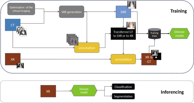
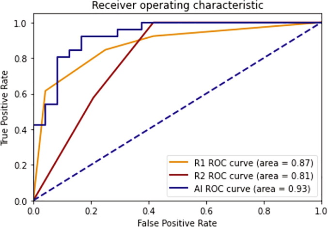
The front-line imaging modalities computed tomography (CT) and X-ray play important roles for triaging COVID patients. Thoracic CT has been accepted to have higher sensitivity than a chest X-ray for COVID diagnosis. Considering the limited access to resources (both hardware and trained personnel) and issues related to decontamination, CT may not be ideal for triaging suspected subjects. Artificial intelligence (AI) assisted X-ray based application for triaging and monitoring require experienced radiologists to identify COVID patients in a timely manner with the additional ability to delineate and quantify the disease region is seen as a promising solution for widespread clinical use. Our proposed solution differs from existing solutions presented by industry and academic communities. We demonstrate a functional AI model to triage by classifying and segmenting a single chest X-ray image, while the AI model is trained using both X-ray and CT data. We report on how such a multi-modal training process improves the solution compared to single modality (X-ray only) training. The multi-modal solution increases the AUC (area under the receiver operating characteristic curve) from 0.89 to 0.93 for a binary classification between COVID-19 and non-COVID-19 cases. It also positively impacts the Dice coefficient (0.59 to 0.62) for localizing the COVID-19 pathology. To compare the performance of experienced readers to the AI model, a reader study is also conducted. The AI model showed good consistency with respect to radiologists. The DICE score between two radiologists on the COVID group was 0.53 while the AI had a DICE value of 0.52 and 0.55 when compared to the segmentation done by the two radiologists separately. From a classification perspective, the AUCs of two readers was 0.87 and 0.81 while the AUC of the AI is 0.93 based on the reader study dataset. We also conducted a generalization study by comparing our method to the-state-art methods on independent datasets. The results show better performance from the proposed method. Leveraging multi-modal information for the development benefits the single-modal inferencing.

摘要

一线成像方式计算机断层扫描(CT)和X射线在对新冠患者进行分流时发挥着重要作用。胸部CT在新冠诊断方面的敏感性已被认为高于胸部X射线。考虑到资源获取有限(包括硬件和训练有素的人员)以及与消毒相关的问题,CT可能并非对疑似病例进行分流的理想选择。基于人工智能(AI)辅助的X射线应用进行分流和监测,需要经验丰富的放射科医生及时识别新冠患者,同时具备描绘和量化疾病区域的额外能力,这被视为广泛临床应用的一个有前景的解决方案。我们提出的解决方案与行业和学术界提出的现有解决方案不同。我们展示了一个通过对单张胸部X射线图像进行分类和分割来进行分流的功能化AI模型,而该AI模型是使用X射线和CT数据进行训练的。我们报告了与单模态(仅X射线)训练相比,这种多模态训练过程如何改进该解决方案。对于新冠病毒感染(COVID - 19)病例和非COVID - 19病例之间的二分类,多模态解决方案将受试者工作特征曲线下面积(AUC)从0.89提高到了0.93。它对定位COVID - 19病变的骰子系数也有积极影响(从0.59提高到0.62)。为了比较经验丰富的阅片者与AI模型的性能,还进行了一项阅片者研究。AI模型与放射科医生表现出良好的一致性。在新冠组中,两位放射科医生之间的骰子系数为0.53,而与两位放射科医生分别进行的分割相比,AI的骰子值为0.52和0.55。从分类角度来看,基于阅片者研究数据集,两位阅片者的AUC分别为0.87和0.81,而AI的AUC为0.93。我们还通过在独立数据集上与最先进的方法进行比较,进行了一项泛化研究。结果表明所提出的方法性能更好。利用多模态信息进行开发有利于单模态推理。

https://cdn.ncbi.nlm.nih.gov/pmc/blobs/ce21/8847079/c7e7d5068c77/gr8_lrg.jpg
https://cdn.ncbi.nlm.nih.gov/pmc/blobs/ce21/8847079/f19a8852d9c6/gr1_lrg.jpg
https://cdn.ncbi.nlm.nih.gov/pmc/blobs/ce21/8847079/29b57db33540/gr2_lrg.jpg
https://cdn.ncbi.nlm.nih.gov/pmc/blobs/ce21/8847079/794d48a97e63/gr3_lrg.jpg
https://cdn.ncbi.nlm.nih.gov/pmc/blobs/ce21/8847079/42d9fb0ed8e4/gr4_lrg.jpg
https://cdn.ncbi.nlm.nih.gov/pmc/blobs/ce21/8847079/8220a75c8d1d/gr5_lrg.jpg
https://cdn.ncbi.nlm.nih.gov/pmc/blobs/ce21/8847079/a0fe0949b57d/gr6_lrg.jpg
https://cdn.ncbi.nlm.nih.gov/pmc/blobs/ce21/8847079/decac620c0c4/gr7_lrg.jpg
https://cdn.ncbi.nlm.nih.gov/pmc/blobs/ce21/8847079/c7e7d5068c77/gr8_lrg.jpg

相似文献

1
Multi-modal trained artificial intelligence solution to triage chest X-ray for COVID-19 using pristine ground-truth, versus radiologists.使用原始真实数据的多模态训练人工智能解决方案与放射科医生对新冠肺炎胸部X光进行分诊。
Neurocomputing (Amst). 2022 May 7;485:36-46. doi: 10.1016/j.neucom.2022.02.040. Epub 2022 Feb 16.
2
From community-acquired pneumonia to COVID-19: a deep learning-based method for quantitative analysis of COVID-19 on thick-section CT scans.从社区获得性肺炎到 COVID-19:一种基于深度学习的 CT 厚层扫描 COVID-19 定量分析方法。
Eur Radiol. 2020 Dec;30(12):6828-6837. doi: 10.1007/s00330-020-07042-x. Epub 2020 Jul 18.
3
Artificial intelligence for segmentation and classification of lobar, lobular, and interstitial pneumonia using case-specific CT information.利用特定病例的CT信息进行大叶性、小叶性和间质性肺炎分割与分类的人工智能技术。
Quant Imaging Med Surg. 2024 Jan 3;14(1):579-591. doi: 10.21037/qims-23-945. Epub 2023 Nov 24.
4
Tuberculosis detection from chest x-rays for triaging in a high tuberculosis-burden setting: an evaluation of five artificial intelligence algorithms.从高结核病负担环境中的胸部 X 光片中检测结核病以进行分诊:五种人工智能算法的评估。
Lancet Digit Health. 2021 Sep;3(9):e543-e554. doi: 10.1016/S2589-7500(21)00116-3.
5
Using artificial intelligence to assist radiologists in distinguishing COVID-19 from other pulmonary infections.利用人工智能辅助放射科医生区分 COVID-19 与其他肺部感染。
J Xray Sci Technol. 2021;29(1):1-17. doi: 10.3233/XST-200735.
6
Is the generalizability of a developed artificial intelligence algorithm for COVID-19 on chest CT sufficient for clinical use? Results from the International Consortium for COVID-19 Imaging AI (ICOVAI).开发用于 COVID-19 胸部 CT 的人工智能算法的可推广性是否足以用于临床?来自 COVID-19 成像人工智能国际联合会(ICOVAI)的结果。
Eur Radiol. 2023 Jun;33(6):4249-4258. doi: 10.1007/s00330-022-09303-3. Epub 2023 Jan 18.
7
AI-based improvement in lung cancer detection on chest radiographs: results of a multi-reader study in NLST dataset.基于人工智能的胸部 X 光肺癌检测改进:NLST 数据集的多读者研究结果。
Eur Radiol. 2021 Dec;31(12):9664-9674. doi: 10.1007/s00330-021-08074-7. Epub 2021 Jun 4.
8
Artificial Intelligence Algorithm Detecting Lung Infection in Supine Chest Radiographs of Critically Ill Patients With a Diagnostic Accuracy Similar to Board-Certified Radiologists.人工智能算法检测重症患者仰卧位胸部 X 光片肺部感染的诊断准确性与放射科认证医师相当。
Crit Care Med. 2020 Jul;48(7):e574-e583. doi: 10.1097/CCM.0000000000004397.
9
Association of Artificial Intelligence-Aided Chest Radiograph Interpretation With Reader Performance and Efficiency.人工智能辅助的胸部 X 光片解读与读者表现和效率的关联。
JAMA Netw Open. 2022 Aug 1;5(8):e2229289. doi: 10.1001/jamanetworkopen.2022.29289.
10
Commercially-available AI algorithm improves radiologists' sensitivity for wrist and hand fracture detection on X-ray, compared to a CT-based ground truth.商业可用的人工智能算法提高了放射科医生在 X 光片上对手腕和手部骨折检测的敏感性,与基于 CT 的金标准相比。
Eur Radiol. 2024 May;34(5):2885-2894. doi: 10.1007/s00330-023-10380-1. Epub 2023 Nov 3.

引用本文的文献

1
A bibliometric analysis of clinical studies on artificial intelligence in emergency medicine.急诊医学中人工智能临床研究的文献计量分析
Medicine (Baltimore). 2025 Jul 11;104(28):e43282. doi: 10.1097/MD.0000000000043282.
2
A Deep Learning Model for the Diagnosis and Discrimination of Gram-Positive and Gram-Negative Bacterial Pneumonia for Children Using Chest Radiography Images and Clinical Information.一种利用胸部X光图像和临床信息对儿童革兰氏阳性和革兰氏阴性细菌性肺炎进行诊断和鉴别的深度学习模型。
Infect Drug Resist. 2023 Jun 24;16:4083-4092. doi: 10.2147/IDR.S404786. eCollection 2023.
3
LightR-YOLOv5: A compact rotating detector for SARS-CoV-2 antigen-detection rapid diagnostic test results.

本文引用的文献

1
Feature Fusion for Diagnosis of Atypical Hepatocellular Carcinoma in Contrast- Enhanced Ultrasound.基于超声造影特征融合的不典型肝细胞癌诊断研究。
IEEE Trans Ultrason Ferroelectr Freq Control. 2022 Jan;69(1):114-123. doi: 10.1109/TUFFC.2021.3110590. Epub 2021 Dec 31.
2
Machine learning applied on chest x-ray can aid in the diagnosis of COVID-19: a first experience from Lombardy, Italy.应用于胸部X光的机器学习有助于COVID-19的诊断:来自意大利伦巴第的首次经验。
Eur Radiol Exp. 2021 Feb 2;5(1):7. doi: 10.1186/s41747-020-00203-z.
3
Hippocampus segmentation in CT using deep learning: impact of MR versus CT-based training contours.
轻量级旋转YOLOv5:用于SARS-CoV-2抗原检测快速诊断测试结果的紧凑型旋转检测器
Displays. 2023 Jul;78:102403. doi: 10.1016/j.displa.2023.102403. Epub 2023 Mar 14.
4
CXray-EffDet: Chest Disease Detection and Classification from X-ray Images Using the EfficientDet Model.CXray-EffDet:使用高效检测(EfficientDet)模型从X光图像中进行胸部疾病检测与分类
Diagnostics (Basel). 2023 Jan 9;13(2):248. doi: 10.3390/diagnostics13020248.
5
Combating Covid-19 using machine learning and deep learning: Applications, challenges, and future perspectives.利用机器学习和深度学习抗击新冠疫情:应用、挑战及未来展望
Array (N Y). 2023 Mar;17:100271. doi: 10.1016/j.array.2022.100271. Epub 2022 Dec 10.
6
Deep fusion of gray level co-occurrence matrices for lung nodule classification.基于灰度共生矩阵的深度融合方法用于肺结节分类。
PLoS One. 2022 Sep 29;17(9):e0274516. doi: 10.1371/journal.pone.0274516. eCollection 2022.
7
AI-CenterNet CXR: An artificial intelligence (AI) enabled system for localization and classification of chest X-ray disease.AI-CenterNet CXR:一种用于胸部X光疾病定位和分类的人工智能(AI)系统。
Front Med (Lausanne). 2022 Aug 30;9:955765. doi: 10.3389/fmed.2022.955765. eCollection 2022.
8
Intubation and mortality prediction in hospitalized COVID-19 patients using a combination of convolutional neural network-based scoring of chest radiographs and clinical data.使用基于卷积神经网络的胸部X光片评分与临床数据相结合的方法对住院COVID-19患者进行插管和死亡率预测
BJR Open. 2022 Mar 24;4(1):20210062. doi: 10.1259/bjro.20210062. eCollection 2022.
基于深度学习的CT图像中海马体分割:基于磁共振成像(MR)与基于CT的训练轮廓的影响
J Med Imaging (Bellingham). 2020 Nov;7(6):064001. doi: 10.1117/1.JMI.7.6.064001. Epub 2020 Nov 11.
4
COVID-Net: a tailored deep convolutional neural network design for detection of COVID-19 cases from chest X-ray images.COVID-Net:一种针对胸部 X 光图像中 COVID-19 病例检测的定制化深度卷积神经网络设计。
Sci Rep. 2020 Nov 11;10(1):19549. doi: 10.1038/s41598-020-76550-z.
5
PadChest: A large chest x-ray image dataset with multi-label annotated reports.PadChest:一个大型胸部 X 射线图像数据集,带有多标签注释报告。
Med Image Anal. 2020 Dec;66:101797. doi: 10.1016/j.media.2020.101797. Epub 2020 Aug 20.
6
Modelling the impact of testing, contact tracing and household quarantine on second waves of COVID-19.建模测试、接触者追踪和家庭隔离对 COVID-19 第二波疫情的影响。
Nat Hum Behav. 2020 Sep;4(9):964-971. doi: 10.1038/s41562-020-0931-9. Epub 2020 Aug 5.
7
Use of Chest Imaging in the Diagnosis and Management of COVID-19: A WHO Rapid Advice Guide.《COVID-19 诊断和管理中胸部成像的应用:世卫组织快速建议指南》。
Radiology. 2021 Feb;298(2):E63-E69. doi: 10.1148/radiol.2020203173. Epub 2020 Jul 30.
8
COVID-19 identification in chest X-ray images on flat and hierarchical classification scenarios.基于平面和分层分类场景的胸部 X 射线图像中的 COVID-19 识别。
Comput Methods Programs Biomed. 2020 Oct;194:105532. doi: 10.1016/j.cmpb.2020.105532. Epub 2020 May 8.
9
Artificial intelligence-enabled rapid diagnosis of patients with COVID-19.人工智能助力 COVID-19 患者快速诊断。
Nat Med. 2020 Aug;26(8):1224-1228. doi: 10.1038/s41591-020-0931-3. Epub 2020 May 19.
10
Variation in False-Negative Rate of Reverse Transcriptase Polymerase Chain Reaction-Based SARS-CoV-2 Tests by Time Since Exposure.基于时间的暴露后,逆转录聚合酶链反应(RT-PCR)检测 SARS-CoV-2 的假阴性率的变化。
Ann Intern Med. 2020 Aug 18;173(4):262-267. doi: 10.7326/M20-1495. Epub 2020 May 13.