• 文献检索
  • 文档翻译
  • 深度研究
  • 学术资讯
  • Suppr Zotero 插件Zotero 插件
  • 邀请有礼
  • 套餐&价格
  • 历史记录
应用&插件
Suppr Zotero 插件Zotero 插件浏览器插件Mac 客户端Windows 客户端微信小程序
定价
高级版会员购买积分包购买API积分包
服务
文献检索文档翻译深度研究API 文档MCP 服务
关于我们
关于 Suppr公司介绍联系我们用户协议隐私条款
关注我们

Suppr 超能文献

核心技术专利:CN118964589B侵权必究
粤ICP备2023148730 号-1Suppr @ 2026

文献检索

告别复杂PubMed语法,用中文像聊天一样搜索,搜遍4000万医学文献。AI智能推荐,让科研检索更轻松。

立即免费搜索

文件翻译

保留排版,准确专业,支持PDF/Word/PPT等文件格式,支持 12+语言互译。

免费翻译文档

深度研究

AI帮你快速写综述,25分钟生成高质量综述,智能提取关键信息,辅助科研写作。

立即免费体验

COVID-Net:一种针对胸部 X 光图像中 COVID-19 病例检测的定制化深度卷积神经网络设计。

COVID-Net: a tailored deep convolutional neural network design for detection of COVID-19 cases from chest X-ray images.

机构信息

Department of Systems Design Engineering, University of Waterloo, Waterloo, Canada.

Waterloo Artificial Intelligence Institute, Waterloo, Canada.

出版信息

Sci Rep. 2020 Nov 11;10(1):19549. doi: 10.1038/s41598-020-76550-z.

DOI:10.1038/s41598-020-76550-z
PMID:33177550
原文链接:https://pmc.ncbi.nlm.nih.gov/articles/PMC7658227/
Abstract

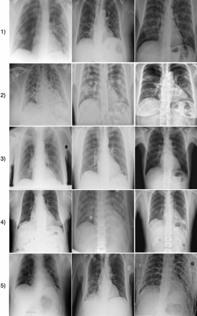
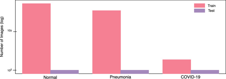
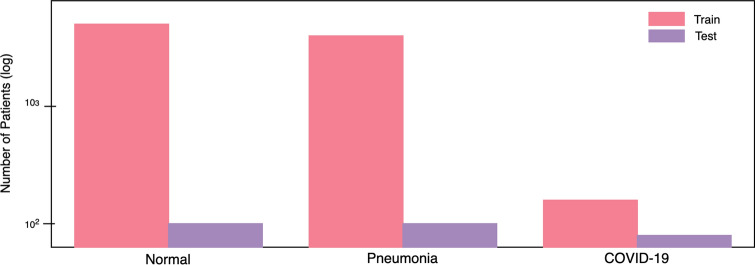
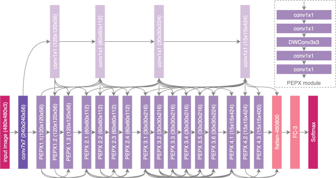
The Coronavirus Disease 2019 (COVID-19) pandemic continues to have a devastating effect on the health and well-being of the global population. A critical step in the fight against COVID-19 is effective screening of infected patients, with one of the key screening approaches being radiology examination using chest radiography. It was found in early studies that patients present abnormalities in chest radiography images that are characteristic of those infected with COVID-19. Motivated by this and inspired by the open source efforts of the research community, in this study we introduce COVID-Net, a deep convolutional neural network design tailored for the detection of COVID-19 cases from chest X-ray (CXR) images that is open source and available to the general public. To the best of the authors' knowledge, COVID-Net is one of the first open source network designs for COVID-19 detection from CXR images at the time of initial release. We also introduce COVIDx, an open access benchmark dataset that we generated comprising of 13,975 CXR images across 13,870 patient patient cases, with the largest number of publicly available COVID-19 positive cases to the best of the authors' knowledge. Furthermore, we investigate how COVID-Net makes predictions using an explainability method in an attempt to not only gain deeper insights into critical factors associated with COVID cases, which can aid clinicians in improved screening, but also audit COVID-Net in a responsible and transparent manner to validate that it is making decisions based on relevant information from the CXR images. By no means a production-ready solution, the hope is that the open access COVID-Net, along with the description on constructing the open source COVIDx dataset, will be leveraged and build upon by both researchers and citizen data scientists alike to accelerate the development of highly accurate yet practical deep learning solutions for detecting COVID-19 cases and accelerate treatment of those who need it the most.

摘要

2019 年冠状病毒病(COVID-19)大流行继续对全球人口的健康和福祉造成破坏性影响。抗击 COVID-19 的关键步骤之一是对感染患者进行有效筛查,其中关键的筛查方法之一是使用胸部 X 射线进行放射学检查。早期研究发现,感染 COVID-19 的患者的胸部 X 射线图像存在异常,这些异常具有特征性。受此启发,并受到研究社区开源工作的启发,在本研究中,我们引入了 COVID-Net,这是一种针对从胸部 X 射线(CXR)图像中检测 COVID-19 病例的深度卷积神经网络设计,该设计是开源的,可供公众使用。据作者所知,COVID-Net 是初始发布时第一个用于从 CXR 图像中检测 COVID-19 的开源网络设计之一。我们还引入了 COVIDx,这是一个开放获取的基准数据集,我们生成了该数据集,其中包含 13870 例患者的 13975 张 CXR 图像,是目前公开的 COVID-19 阳性病例数量最多的数据集。此外,我们还研究了 COVID-Net 如何使用可解释性方法进行预测,以便不仅深入了解与 COVID 病例相关的关键因素,从而帮助临床医生进行更好的筛查,而且还以负责任和透明的方式对 COVID-Net 进行审核,以验证它是根据 CXR 图像中的相关信息做出决策的。绝不是一种可立即投入使用的解决方案,我们希望开放访问的 COVID-Net 以及构建开源 COVIDx 数据集的说明,将被研究人员和公民数据科学家利用和进一步开发,以加速开发高度准确但实用的深度学习解决方案,用于检测 COVID-19 病例,并加速对最需要的患者的治疗。

https://cdn.ncbi.nlm.nih.gov/pmc/blobs/f2d7/7658227/569caed5da88/41598_2020_76550_Fig7_HTML.jpg
https://cdn.ncbi.nlm.nih.gov/pmc/blobs/f2d7/7658227/c1acf3b2e352/41598_2020_76550_Fig1_HTML.jpg
https://cdn.ncbi.nlm.nih.gov/pmc/blobs/f2d7/7658227/47fac028ae0b/41598_2020_76550_Fig2_HTML.jpg
https://cdn.ncbi.nlm.nih.gov/pmc/blobs/f2d7/7658227/1ed4f9ab5dc2/41598_2020_76550_Fig3_HTML.jpg
https://cdn.ncbi.nlm.nih.gov/pmc/blobs/f2d7/7658227/e703a542fcab/41598_2020_76550_Fig4_HTML.jpg
https://cdn.ncbi.nlm.nih.gov/pmc/blobs/f2d7/7658227/65f443064d27/41598_2020_76550_Fig5_HTML.jpg
https://cdn.ncbi.nlm.nih.gov/pmc/blobs/f2d7/7658227/00a3f5eca416/41598_2020_76550_Fig6_HTML.jpg
https://cdn.ncbi.nlm.nih.gov/pmc/blobs/f2d7/7658227/569caed5da88/41598_2020_76550_Fig7_HTML.jpg

相似文献

1
COVID-Net: a tailored deep convolutional neural network design for detection of COVID-19 cases from chest X-ray images.COVID-Net:一种针对胸部 X 光图像中 COVID-19 病例检测的定制化深度卷积神经网络设计。
Sci Rep. 2020 Nov 11;10(1):19549. doi: 10.1038/s41598-020-76550-z.
2
COVIDNet-CT: A Tailored Deep Convolutional Neural Network Design for Detection of COVID-19 Cases From Chest CT Images.COVIDNet-CT:一种用于从胸部CT图像中检测新冠肺炎病例的定制深度卷积神经网络设计。
Front Med (Lausanne). 2020 Dec 23;7:608525. doi: 10.3389/fmed.2020.608525. eCollection 2020.
3
COVID-Net CT-2: Enhanced Deep Neural Networks for Detection of COVID-19 From Chest CT Images Through Bigger, More Diverse Learning.COVID-Net CT-2:通过更大规模、更多样化的学习从胸部CT图像中检测新型冠状病毒肺炎的增强深度神经网络
Front Med (Lausanne). 2022 Mar 10;8:729287. doi: 10.3389/fmed.2021.729287. eCollection 2021.
4
COVID-Net CXR-2: An Enhanced Deep Convolutional Neural Network Design for Detection of COVID-19 Cases From Chest X-ray Images.COVID-Net CXR-2:一种用于从胸部X光图像中检测新冠肺炎病例的增强型深度卷积神经网络设计。
Front Med (Lausanne). 2022 Jun 10;9:861680. doi: 10.3389/fmed.2022.861680. eCollection 2022.
5
Identification of COVID-19 samples from chest X-Ray images using deep learning: A comparison of transfer learning approaches.基于深度学习的胸部 X 光图像中 COVID-19 样本的识别:迁移学习方法的比较。
J Xray Sci Technol. 2020;28(5):821-839. doi: 10.3233/XST-200715.
6
TB-Net: A Tailored, Self-Attention Deep Convolutional Neural Network Design for Detection of Tuberculosis Cases From Chest X-Ray Images.TB-Net:一种用于从胸部X光图像中检测肺结核病例的定制化自注意力深度卷积神经网络设计。
Front Artif Intell. 2022 Apr 7;5:827299. doi: 10.3389/frai.2022.827299. eCollection 2022.
7
CoroNet: A deep neural network for detection and diagnosis of COVID-19 from chest x-ray images.CoroNet:一种用于从胸部 X 光图像中检测和诊断 COVID-19 的深度神经网络。
Comput Methods Programs Biomed. 2020 Nov;196:105581. doi: 10.1016/j.cmpb.2020.105581. Epub 2020 Jun 5.
8
Deep-COVID: Predicting COVID-19 from chest X-ray images using deep transfer learning.深度 COVID:使用深度迁移学习从胸部 X 光图像预测 COVID-19。
Med Image Anal. 2020 Oct;65:101794. doi: 10.1016/j.media.2020.101794. Epub 2020 Jul 21.
9
Cancer-Net SCa: tailored deep neural network designs for detection of skin cancer from dermoscopy images.Cancer-Net SCa:用于从皮肤镜图像中检测皮肤癌的定制深度神经网络设计。
BMC Med Imaging. 2022 Aug 9;22(1):143. doi: 10.1186/s12880-022-00871-w.
10
CovXNet: A multi-dilation convolutional neural network for automatic COVID-19 and other pneumonia detection from chest X-ray images with transferable multi-receptive feature optimization.CovXNet:一种多扩张卷积神经网络,用于从胸部 X 光图像中自动检测 COVID-19 和其他肺炎,具有可转移的多感受野特征优化。
Comput Biol Med. 2020 Jul;122:103869. doi: 10.1016/j.compbiomed.2020.103869. Epub 2020 Jun 20.

引用本文的文献

1
Measuring Subtle HD Data Representation and Multimodal Imaging Phenotype Embedding for Precision Medicine.测量用于精准医学的细微高清数据表征和多模态成像表型嵌入
IEEE Trans Instrum Meas. 2025;74. doi: 10.1109/tim.2025.3545983. Epub 2025 Mar 3.
2
Improving CNN predictive accuracy in COVID-19 health analytics.提高新冠疫情健康分析中卷积神经网络的预测准确性。
Sci Rep. 2025 Aug 14;15(1):29864. doi: 10.1038/s41598-025-15218-y.
3
Segmentation-Assisted Fusion-Based Classification for Automated CXR Image Analysis.基于分割辅助融合的自动胸部X光图像分析分类方法

本文引用的文献

1
Imaging Profile of the COVID-19 Infection: Radiologic Findings and Literature Review.新型冠状病毒肺炎感染的影像学表现:放射学发现与文献综述
Radiol Cardiothorac Imaging. 2020 Feb 13;2(1):e200034. doi: 10.1148/ryct.2020200034. eCollection 2020 Feb.
2
Large-scale screening to distinguish between COVID-19 and community-acquired pneumonia using infection size-aware classification.利用感染大小感知分类进行大规模筛选,以区分 COVID-19 和社区获得性肺炎。
Phys Med Biol. 2021 Mar 17;66(6):065031. doi: 10.1088/1361-6560/abe838.
3
Estimating the false-negative test probability of SARS-CoV-2 by RT-PCR.
Sensors (Basel). 2025 Jul 24;25(15):4580. doi: 10.3390/s25154580.
4
Reassessing deep learning (and meta-learning) computer vision as an efficient method to determine taphonomic agency in bone surface modifications.重新评估深度学习(和元学习)计算机视觉作为确定骨表面改变中埋藏作用的一种有效方法。
Biol Methods Protoc. 2025 Jul 12;10(1):bpaf057. doi: 10.1093/biomethods/bpaf057. eCollection 2025.
5
Differentiation of COVID-19 from other types of viral pneumonia and severity scoring on baseline chest radiographs: Comparison of deep learning with multi-reader evaluation.基于胸部X光片将新型冠状病毒肺炎与其他类型病毒性肺炎进行鉴别及严重程度评分:深度学习与多位阅片者评估的比较
PLoS One. 2025 Jul 29;20(7):e0328061. doi: 10.1371/journal.pone.0328061. eCollection 2025.
6
Classification of Pneumonia, Tuberculosis and Covid-19 from Chest X-Ray Images Using Convolution Neural Network Model.使用卷积神经网络模型从胸部X光图像中对肺炎、肺结核和新冠肺炎进行分类
Int J Stat Probab. 2024;13(4):42-63. doi: 10.5539/ijsp.v13n4p42. Epub 2024 Nov 30.
7
Enhancing Pandemic Prediction: A Deep Learning Approach Using Transformer Neural Networks and Multi-Source Data Fusion for Infectious Disease Forecasting.增强大流行预测:一种使用Transformer神经网络和多源数据融合进行传染病预测的深度学习方法。
medRxiv. 2025 Jun 24:2025.06.24.25330211. doi: 10.1101/2025.06.24.25330211.
8
Leveraging federated learning and edge computing for pandemic-resilient healthcare.利用联邦学习和边缘计算实现抗疫情的医疗保健。
Sci Rep. 2025 Jul 1;15(1):20497. doi: 10.1038/s41598-025-00199-9.
9
Parallel VMamba and Attention-Based Pneumonia Severity Prediction from CXRs: A Robust Model with Segmented Lung Replacement Augmentation.基于并行VMamba和注意力机制的胸部X光片肺炎严重程度预测:一种采用分割肺替代增强的稳健模型
Diagnostics (Basel). 2025 May 22;15(11):1301. doi: 10.3390/diagnostics15111301.
10
A fully open AI foundation model applied to chest radiography.一种应用于胸部X光摄影的完全开放的人工智能基础模型。
Nature. 2025 Jun 11. doi: 10.1038/s41586-025-09079-8.
估算 RT-PCR 检测 SARS-CoV-2 的假阴性率。
Euro Surveill. 2020 Dec;25(50). doi: 10.2807/1560-7917.ES.2020.25.50.2000568.
4
Unveiling COVID-19 from CHEST X-Ray with Deep Learning: A Hurdles Race with Small Data.利用深度学习揭示 CHEST X-RAY 中的 COVID-19:小数据的障碍竞赛。
Int J Environ Res Public Health. 2020 Sep 22;17(18):6933. doi: 10.3390/ijerph17186933.
5
COVID-CAPS: A capsule network-based framework for identification of COVID-19 cases from X-ray images.COVID-CAPS:一种基于胶囊网络的从X射线图像识别新冠肺炎病例的框架。
Pattern Recognit Lett. 2020 Oct;138:638-643. doi: 10.1016/j.patrec.2020.09.010. Epub 2020 Sep 16.
6
Deep-COVID: Predicting COVID-19 from chest X-ray images using deep transfer learning.深度 COVID:使用深度迁移学习从胸部 X 光图像预测 COVID-19。
Med Image Anal. 2020 Oct;65:101794. doi: 10.1016/j.media.2020.101794. Epub 2020 Jul 21.
7
Covid-19: automatic detection from X-ray images utilizing transfer learning with convolutional neural networks.新冠病毒(Covid-19):利用卷积神经网络的迁移学习从 X 光图像中自动检测。
Phys Eng Sci Med. 2020 Jun;43(2):635-640. doi: 10.1007/s13246-020-00865-4. Epub 2020 Apr 3.
8
Assessing risk factors for SARS-CoV-2 infection in patients presenting with symptoms in Shanghai, China: a multicentre, observational cohort study.评估中国上海出现症状的患者感染 SARS-CoV-2 的风险因素:一项多中心、观察性队列研究。
Lancet Digit Health. 2020 Jun;2(6):e323-e330. doi: 10.1016/S2589-7500(20)30109-6. Epub 2020 May 14.
9
Mobile X-rays are highly valuable for critically ill COVID patients.移动X光机对危重症新冠患者非常有价值。
Eur Radiol. 2020 Sep;30(9):5217-5219. doi: 10.1007/s00330-020-06918-2. Epub 2020 May 13.
10
Canadian Society of Thoracic Radiology/Canadian Association of Radiologists Consensus Statement Regarding Chest Imaging in Suspected and Confirmed COVID-19.加拿大胸放射学会/加拿大放射学家协会关于疑似和确诊 COVID-19 胸部成像的共识声明。
Can Assoc Radiol J. 2020 Nov;71(4):470-481. doi: 10.1177/0846537120924606. Epub 2020 May 8.