• 文献检索
  • 文档翻译
  • 深度研究
  • 学术资讯
  • Suppr Zotero 插件Zotero 插件
  • 邀请有礼
  • 套餐&价格
  • 历史记录
应用&插件
Suppr Zotero 插件Zotero 插件浏览器插件Mac 客户端Windows 客户端微信小程序
定价
高级版会员购买积分包购买API积分包
服务
文献检索文档翻译深度研究API 文档MCP 服务
关于我们
关于 Suppr公司介绍联系我们用户协议隐私条款
关注我们

Suppr 超能文献

核心技术专利:CN118964589B侵权必究
粤ICP备2023148730 号-1Suppr @ 2026

文献检索

告别复杂PubMed语法,用中文像聊天一样搜索,搜遍4000万医学文献。AI智能推荐,让科研检索更轻松。

立即免费搜索

文件翻译

保留排版,准确专业,支持PDF/Word/PPT等文件格式,支持 12+语言互译。

免费翻译文档

深度研究

AI帮你快速写综述,25分钟生成高质量综述,智能提取关键信息,辅助科研写作。

立即免费体验

基于平面和分层分类场景的胸部 X 射线图像中的 COVID-19 识别。

COVID-19 identification in chest X-ray images on flat and hierarchical classification scenarios.

机构信息

Instituto Federal de Educação, Ciência e Tecnologia do Paraná (IFPR), Pinhais, PR, Brazil; Pontifícia Universidade Catalica do Paraná (PUCPR), Curitiba, PR, Brazil.

Universidade Tecnologica Federal do Paraná (UTFPR), Campo Mourão, PR, Brazil; Universidade Estadual de Maringá (UEM), Maringá, PR, Brazil.

出版信息

Comput Methods Programs Biomed. 2020 Oct;194:105532. doi: 10.1016/j.cmpb.2020.105532. Epub 2020 May 8.

DOI:10.1016/j.cmpb.2020.105532
PMID:32446037
原文链接:https://pmc.ncbi.nlm.nih.gov/articles/PMC7207172/
Abstract

BACKGROUND AND OBJECTIVE

The COVID-19 can cause severe pneumonia and is estimated to have a high impact on the healthcare system. Early diagnosis is crucial for correct treatment in order to possibly reduce the stress in the healthcare system. The standard image diagnosis tests for pneumonia are chest X-ray (CXR) and computed tomography (CT) scan. Although CT scan is the gold standard, CXR are still useful because it is cheaper, faster and more widespread. This study aims to identify pneumonia caused by COVID-19 from other types and also healthy lungs using only CXR images.

METHODS

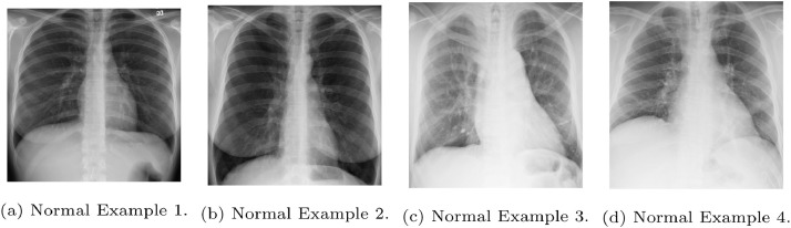
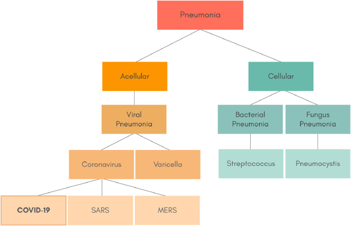
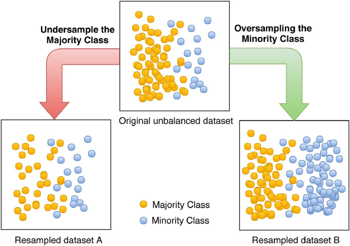
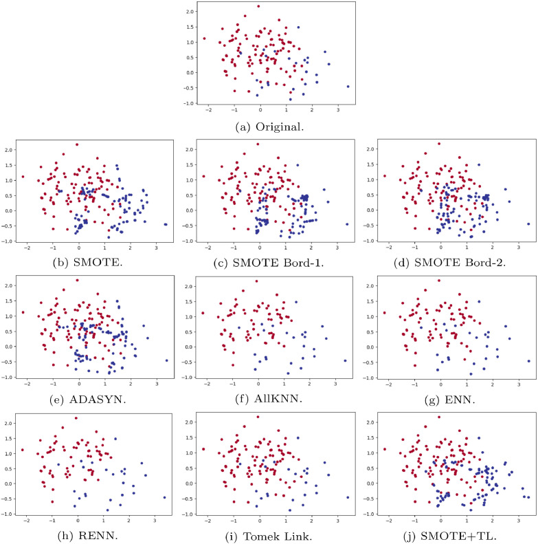
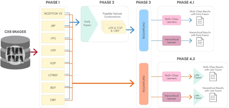
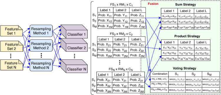
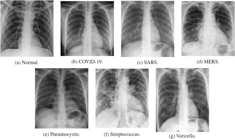
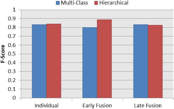
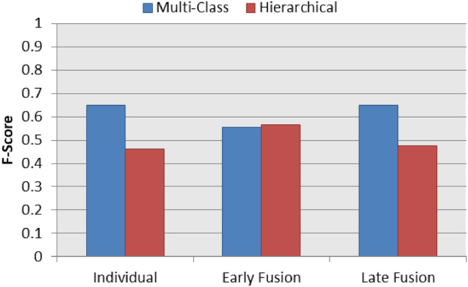
In order to achieve the objectives, we have proposed a classification schema considering the following perspectives: i) a multi-class classification; ii) hierarchical classification, since pneumonia can be structured as a hierarchy. Given the natural data imbalance in this domain, we also proposed the use of resampling algorithms in the schema in order to re-balance the classes distribution. We observed that, texture is one of the main visual attributes of CXR images, our classification schema extract features using some well-known texture descriptors and also using a pre-trained CNN model. We also explored early and late fusion techniques in the schema in order to leverage the strength of multiple texture descriptors and base classifiers at once. To evaluate the approach, we composed a database, named RYDLS-20, containing CXR images of pneumonia caused by different pathogens as well as CXR images of healthy lungs. The classes distribution follows a real-world scenario in which some pathogens are more common than others.

RESULTS

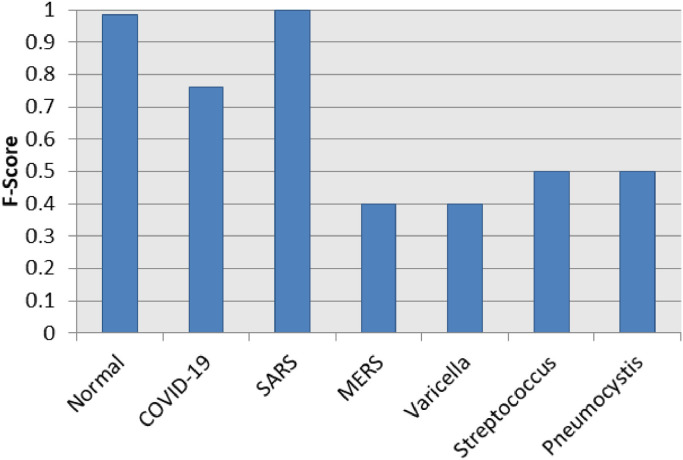
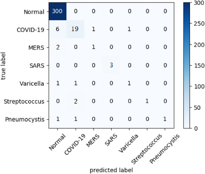
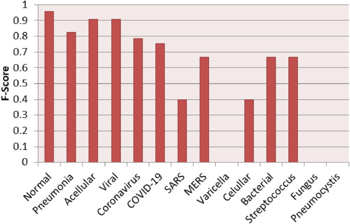
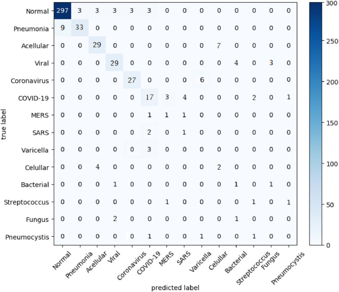
The proposed approach tested in RYDLS-20 achieved a macro-avg F1-Score of 0.65 using a multi-class approach and a F1-Score of 0.89 for the COVID-19 identification in the hierarchical classification scenario.

CONCLUSIONS

As far as we know, the top identification rate obtained in this paper is the best nominal rate obtained for COVID-19 identification in an unbalanced environment with more than three classes. We must also highlight the novel proposed hierarchical classification approach for this task, which considers the types of pneumonia caused by the different pathogens and lead us to the best COVID-19 recognition rate obtained here.

摘要

背景与目的

新冠病毒可引起严重肺炎,预计对医疗体系有较大影响。早期诊断对正确治疗至关重要,以便可能减轻医疗体系的压力。肺炎的标准影像学诊断检查为胸部 X 光(CXR)和计算机断层扫描(CT)。尽管 CT 扫描是金标准,但 CXR 仍具有价值,因为其更便宜、更快且更普及。本研究旨在仅使用 CXR 图像,从其他类型肺炎和健康肺中识别出由新冠病毒引起的肺炎。

方法

为了实现目标,我们提出了一种分类方案,考虑了以下方面:i)多类分类;ii)层次分类,因为肺炎可以分层结构。鉴于该领域数据天然不平衡,我们还提出在方案中使用重采样算法来重新平衡类别的分布。我们观察到,纹理是 CXR 图像的主要视觉属性之一,我们的分类方案使用一些知名的纹理描述符以及预训练的 CNN 模型来提取特征。我们还在方案中探索了早期和晚期融合技术,以一次利用多个纹理描述符和基础分类器的优势。为了评估该方法,我们构建了一个名为 RYDLS-20 的数据库,其中包含了由不同病原体引起的肺炎和健康肺的 CXR 图像。类别的分布遵循实际情况,即一些病原体比其他病原体更常见。

结果

在 RYDLS-20 中测试的所提出的方法在多类方法中实现了 0.65 的宏平均 F1-分数,在层次分类场景中实现了 0.89 的 COVID-19 识别 F1-分数。

结论

据我们所知,本文获得的最高识别率是在具有三类以上的不平衡环境中对 COVID-19 识别获得的最佳标称率。我们还必须强调为此任务提出的新颖层次分类方法,它考虑了由不同病原体引起的不同类型肺炎,并导致我们在这里获得了最佳的 COVID-19 识别率。

https://cdn.ncbi.nlm.nih.gov/pmc/blobs/7b77/7207172/f6046099df17/gr14_lrg.jpg
https://cdn.ncbi.nlm.nih.gov/pmc/blobs/7b77/7207172/f0998140f322/fx1_lrg.jpg
https://cdn.ncbi.nlm.nih.gov/pmc/blobs/7b77/7207172/085584f72248/gr1_lrg.jpg
https://cdn.ncbi.nlm.nih.gov/pmc/blobs/7b77/7207172/5a61a2079c92/gr2_lrg.jpg
https://cdn.ncbi.nlm.nih.gov/pmc/blobs/7b77/7207172/76b22e2bc9df/gr3_lrg.jpg
https://cdn.ncbi.nlm.nih.gov/pmc/blobs/7b77/7207172/d39f8f072a28/gr4_lrg.jpg
https://cdn.ncbi.nlm.nih.gov/pmc/blobs/7b77/7207172/8e06bcaa8ba9/gr5_lrg.jpg
https://cdn.ncbi.nlm.nih.gov/pmc/blobs/7b77/7207172/71de40ee2e7e/gr6_lrg.jpg
https://cdn.ncbi.nlm.nih.gov/pmc/blobs/7b77/7207172/34c268a004b5/gr7_lrg.jpg
https://cdn.ncbi.nlm.nih.gov/pmc/blobs/7b77/7207172/60c49e35b8b6/gr8_lrg.jpg
https://cdn.ncbi.nlm.nih.gov/pmc/blobs/7b77/7207172/86cd06d4f7b7/gr9_lrg.jpg
https://cdn.ncbi.nlm.nih.gov/pmc/blobs/7b77/7207172/1aa5bd774583/gr10_lrg.jpg
https://cdn.ncbi.nlm.nih.gov/pmc/blobs/7b77/7207172/aaa8a4c67c70/gr11_lrg.jpg
https://cdn.ncbi.nlm.nih.gov/pmc/blobs/7b77/7207172/16247d3e5be2/gr12_lrg.jpg
https://cdn.ncbi.nlm.nih.gov/pmc/blobs/7b77/7207172/544983d72de0/gr13_lrg.jpg
https://cdn.ncbi.nlm.nih.gov/pmc/blobs/7b77/7207172/f6046099df17/gr14_lrg.jpg

相似文献

1
COVID-19 identification in chest X-ray images on flat and hierarchical classification scenarios.基于平面和分层分类场景的胸部 X 射线图像中的 COVID-19 识别。
Comput Methods Programs Biomed. 2020 Oct;194:105532. doi: 10.1016/j.cmpb.2020.105532. Epub 2020 May 8.
2
CovXNet: A multi-dilation convolutional neural network for automatic COVID-19 and other pneumonia detection from chest X-ray images with transferable multi-receptive feature optimization.CovXNet:一种多扩张卷积神经网络,用于从胸部 X 光图像中自动检测 COVID-19 和其他肺炎,具有可转移的多感受野特征优化。
Comput Biol Med. 2020 Jul;122:103869. doi: 10.1016/j.compbiomed.2020.103869. Epub 2020 Jun 20.
3
Identification of COVID-19 samples from chest X-Ray images using deep learning: A comparison of transfer learning approaches.基于深度学习的胸部 X 光图像中 COVID-19 样本的识别:迁移学习方法的比较。
J Xray Sci Technol. 2020;28(5):821-839. doi: 10.3233/XST-200715.
4
CoroNet: A deep neural network for detection and diagnosis of COVID-19 from chest x-ray images.CoroNet:一种用于从胸部 X 光图像中检测和诊断 COVID-19 的深度神经网络。
Comput Methods Programs Biomed. 2020 Nov;196:105581. doi: 10.1016/j.cmpb.2020.105581. Epub 2020 Jun 5.
5
Adaptive Feature Selection Guided Deep Forest for COVID-19 Classification With Chest CT.基于胸部 CT 的 COVID-19 分类的自适应特征选择引导深度森林
IEEE J Biomed Health Inform. 2020 Oct;24(10):2798-2805. doi: 10.1109/JBHI.2020.3019505. Epub 2020 Aug 26.
6
Thoracic imaging tests for the diagnosis of COVID-19.用于诊断新型冠状病毒肺炎的胸部影像学检查
Cochrane Database Syst Rev. 2020 Sep 30;9:CD013639. doi: 10.1002/14651858.CD013639.pub2.
7
COVID19XrayNet: A Two-Step Transfer Learning Model for the COVID-19 Detecting Problem Based on a Limited Number of Chest X-Ray Images.COVID19XrayNet:一种基于少量胸部 X 光图像的 COVID-19 检测问题的两步迁移学习模型。
Interdiscip Sci. 2020 Dec;12(4):555-565. doi: 10.1007/s12539-020-00393-5. Epub 2020 Sep 21.
8
Classification of COVID-19 patients from chest CT images using multi-objective differential evolution-based convolutional neural networks.基于多目标差分进化卷积神经网络的胸部 CT 图像 COVID-19 患者分类。
Eur J Clin Microbiol Infect Dis. 2020 Jul;39(7):1379-1389. doi: 10.1007/s10096-020-03901-z. Epub 2020 Apr 27.
9
COVID-19 Hierarchical Classification Using a Deep Learning Multi-Modal.使用深度学习多模态的COVID-19分层分类
Sensors (Basel). 2024 Apr 20;24(8):2641. doi: 10.3390/s24082641.
10
Deep Learning COVID-19 Features on CXR Using Limited Training Data Sets.基于有限训练数据集的 X 光胸片深度学习 COVID-19 特征
IEEE Trans Med Imaging. 2020 Aug;39(8):2688-2700. doi: 10.1109/TMI.2020.2993291. Epub 2020 May 8.

引用本文的文献

1
Deep Learning Network Selection and Optimized Information Fusion for Enhanced COVID-19 Detection: A Literature Review.用于增强COVID-19检测的深度学习网络选择与优化信息融合:文献综述
Diagnostics (Basel). 2025 Jul 21;15(14):1830. doi: 10.3390/diagnostics15141830.
2
Deep learning-based cough classification using application-recorded sounds: a transfer learning approach with VGGish.基于深度学习的咳嗽分类:利用应用程序录制声音,采用VGGish的迁移学习方法
BMC Med Inform Decis Mak. 2025 Jul 1;25(1):228. doi: 10.1186/s12911-025-03065-w.
3
A novel normalization algorithm to facilitate pre-assessment of Covid-19 disease by improving accuracy of CNN and its FPGA implementation.

本文引用的文献

1
Automatic detection of coronavirus disease (COVID-19) using X-ray images and deep convolutional neural networks.使用X射线图像和深度卷积神经网络自动检测冠状病毒病(COVID-19)。
Pattern Anal Appl. 2021;24(3):1207-1220. doi: 10.1007/s10044-021-00984-y. Epub 2021 May 9.
2
A critic evaluation of methods for COVID-19 automatic detection from X-ray images.对从X射线图像中自动检测COVID-19的方法的批判性评估。
Inf Fusion. 2021 Dec;76:1-7. doi: 10.1016/j.inffus.2021.04.008. Epub 2021 Apr 30.
3
Deep learning for differentiating novel coronavirus pneumonia and influenza pneumonia.
一种通过提高卷积神经网络(CNN)的准确性来促进新冠病毒疾病预评估的新型归一化算法及其现场可编程门阵列(FPGA)实现。
Evol Syst (Berl). 2022 Feb 1:1-11. doi: 10.1007/s12530-022-09419-3.
4
Deep Learning for Pneumonia Detection in Chest X-ray Images: A Comprehensive Survey.胸部X光图像中肺炎检测的深度学习:全面综述。
J Imaging. 2024 Jul 23;10(8):176. doi: 10.3390/jimaging10080176.
5
Adaptive Mish activation and ranger optimizer-based SEA-ResNet50 model with explainable AI for multiclass classification of COVID-19 chest X-ray images.基于自适应 Mish 激活和 Ranger 优化器的 SEA-ResNet50 模型,具有可解释 AI,用于 COVID-19 胸部 X 射线图像的多类分类。
BMC Med Imaging. 2024 Aug 9;24(1):206. doi: 10.1186/s12880-024-01394-2.
6
Optimization of vision transformer-based detection of lung diseases from chest X-ray images.基于视觉Transformer的胸部 X 射线图像肺病检测的优化。
BMC Med Inform Decis Mak. 2024 Jul 8;24(1):191. doi: 10.1186/s12911-024-02591-3.
7
TransMVAN: Multi-view Aggregation Network with Transformer for Pneumonia Diagnosis.TransMVAN:用于肺炎诊断的基于Transformer的多视图聚合网络。
J Imaging Inform Med. 2025 Feb;38(1):60-73. doi: 10.1007/s10278-024-01169-9. Epub 2024 Jul 8.
8
A machine learning approach using conditional normalizing flow to address extreme class imbalance problems in personal health records.一种使用条件归一化流来解决个人健康记录中极端类别不平衡问题的机器学习方法。
BioData Min. 2024 May 25;17(1):14. doi: 10.1186/s13040-024-00366-0.
9
COVID-19 Hierarchical Classification Using a Deep Learning Multi-Modal.使用深度学习多模态的COVID-19分层分类
Sensors (Basel). 2024 Apr 20;24(8):2641. doi: 10.3390/s24082641.
10
AMTLDC: a new adversarial multi-source transfer learning framework to diagnosis of COVID-19.AMTLDC:一种用于新冠肺炎诊断的新型对抗多源迁移学习框架。
Evol Syst (Berl). 2023 Jan 12:1-15. doi: 10.1007/s12530-023-09484-2.
用于区分新型冠状病毒肺炎和流感肺炎的深度学习
Ann Transl Med. 2021 Jan;9(2):111. doi: 10.21037/atm-20-5328.
4
COVID-Net: a tailored deep convolutional neural network design for detection of COVID-19 cases from chest X-ray images.COVID-Net:一种针对胸部 X 光图像中 COVID-19 病例检测的定制化深度卷积神经网络设计。
Sci Rep. 2020 Nov 11;10(1):19549. doi: 10.1038/s41598-020-76550-z.
5
Automated detection of COVID-19 cases using deep neural networks with X-ray images.使用 X 射线图像的深度学习神经网络自动检测 COVID-19 病例。
Comput Biol Med. 2020 Jun;121:103792. doi: 10.1016/j.compbiomed.2020.103792. Epub 2020 Apr 28.
6
CoroNet: A deep neural network for detection and diagnosis of COVID-19 from chest x-ray images.CoroNet:一种用于从胸部 X 光图像中检测和诊断 COVID-19 的深度神经网络。
Comput Methods Programs Biomed. 2020 Nov;196:105581. doi: 10.1016/j.cmpb.2020.105581. Epub 2020 Jun 5.
7
The Role of Chest Imaging in Patient Management During the COVID-19 Pandemic: A Multinational Consensus Statement From the Fleischner Society.COVID-19 大流行期间患者管理中的胸部影像学作用: Fleischner 学会的一项多国家共识声明。
Chest. 2020 Jul;158(1):106-116. doi: 10.1016/j.chest.2020.04.003. Epub 2020 Apr 7.
8
Using Artificial Intelligence to Detect COVID-19 and Community-acquired Pneumonia Based on Pulmonary CT: Evaluation of the Diagnostic Accuracy.基于肺部 CT 的人工智能检测 COVID-19 和社区获得性肺炎:诊断准确性评估。
Radiology. 2020 Aug;296(2):E65-E71. doi: 10.1148/radiol.2020200905. Epub 2020 Mar 19.
9
Influenza-associated pneumonia as reference to assess seriousness of coronavirus disease (COVID-19).将流感相关性肺炎作为参考来评估新型冠状病毒病(COVID-19)的严重程度。
Euro Surveill. 2020 Mar;25(11). doi: 10.2807/1560-7917.ES.2020.25.11.2000258. Epub 2020 Mar 16.
10
COVID-19 and Italy: what next?COVID-19 和意大利:下一步如何?
Lancet. 2020 Apr 11;395(10231):1225-1228. doi: 10.1016/S0140-6736(20)30627-9. Epub 2020 Mar 13.