Ho Kai-Yu, Liang Jing Nong, Budge Savanna, Madriaga Austin, Meske Kara, Nguyenton Derrick
Department of Physical Therapy, University of Nevada, Las Vegas, Las Vegas, NV, United States.
Front Integr Neurosci. 2022 Feb 7;16:791719. doi: 10.3389/fnint.2022.791719. eCollection 2022.
To evaluate the evidence for altered cortical and spinal cord functions in individuals with patellofemoral pain (PFP).
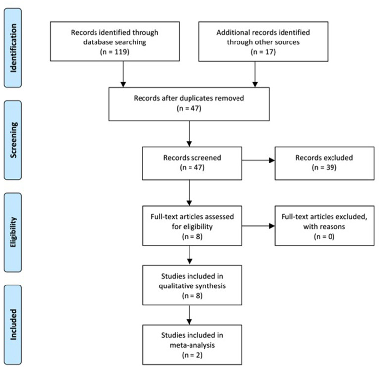
We conducted a comprehensive search of databases to appraise and analyze the studies published prior to December 10, 2021 that examined spinal reflex excitability measured using Hoffmann reflex (H-reflex) amplitudes, corticospinal excitability measured using transcranial magnetic stimulation (TMS)-elicited motor evoked potential (MEP) amplitudes, motor threshold (MT), or stimulus-response (SR) curves, cortical reorganization assessed using TMS cortical mapping or structural magnetic resonance imaging (MRI), or functional changes of the brain assessed using functional MRI (fMRI) in individuals with PFP.
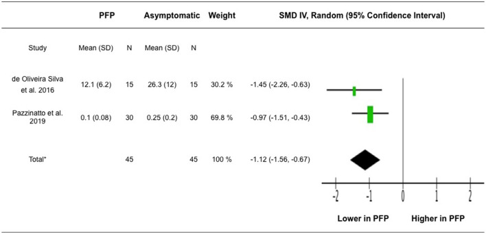
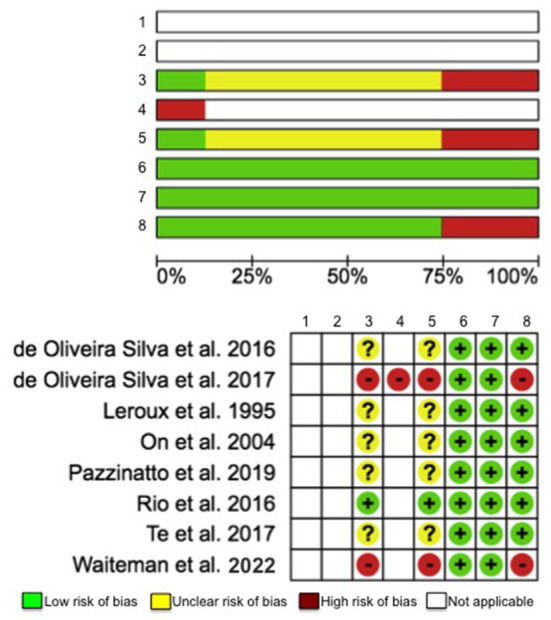
Eight studies were eligible for analyses. While an earlier study showed that pain had no effect on the H-reflex amplitude of the quadriceps muscle, more recent evidence reported a decrease in vastus medialis (VM) H-reflex amplitude in participants with PFP. VM H-reflex amplitude was correlated with pain, chronicity, physical function, and isometric knee extensor torque production in participants with PFP. Altered corticospinal excitability was reported in participants with PFP, observed as increased MT in the VM and vastus lateralis (VL) muscles. In addition, cortical reorganization has been observed, where decreased number of cortical peaks, shifts and reduced volumes, and increased overlap of motor cortex representations for the VM, VL, and rectus femoris (RF) muscles were reported in participants with PFP.
There is emerging evidence on altered cortical and spinal cord functions in individuals with PFP, however, solid conclusions cannot be drawn due to limited literature available. Further research is needed to better understand the adaptations of the brain and spinal cord in this population.
https://www.crd.york.ac.uk/prospero/, identifier: CRD42020212128.
评估髌股疼痛(PFP)患者皮质和脊髓功能改变的证据。
我们对数据库进行了全面检索,以评估和分析2021年12月10日前发表的研究,这些研究检测了使用霍夫曼反射(H反射)幅度测量的脊髓反射兴奋性、使用经颅磁刺激(TMS)诱发的运动诱发电位(MEP)幅度、运动阈值(MT)或刺激-反应(SR)曲线测量的皮质脊髓兴奋性、使用TMS皮质映射或结构磁共振成像(MRI)评估的皮质重组,或使用功能磁共振成像(fMRI)评估的PFP患者大脑的功能变化。
八项研究符合分析条件。虽然一项早期研究表明疼痛对股四头肌的H反射幅度没有影响,但最近的证据报告称,PFP患者的股内侧肌(VM)H反射幅度降低。VM H反射幅度与PFP患者的疼痛、病程、身体功能和等长膝关节伸肌扭矩产生相关。PFP患者报告有皮质脊髓兴奋性改变,表现为VM和股外侧肌(VL)肌肉的MT增加。此外,还观察到皮质重组,PFP患者报告VM、VL和股直肌(RF)肌肉的皮质峰值数量减少、移位和体积减小,以及运动皮质表征的重叠增加。
有新证据表明PFP患者存在皮质和脊髓功能改变,然而,由于现有文献有限,无法得出确凿结论。需要进一步研究以更好地了解该人群大脑和脊髓的适应性变化。