The Sidney Kimmel Comprehensive Cancer Center, Department of Oncology, The Johns Hopkins University School of Medicine, Baltimore, MD 21231, USA.
Department of Chemical and Biomolecular Engineering, The Johns Hopkins University, Baltimore, MD 21218, USA.
Cells. 2022 Feb 16;11(4):686. doi: 10.3390/cells11040686.
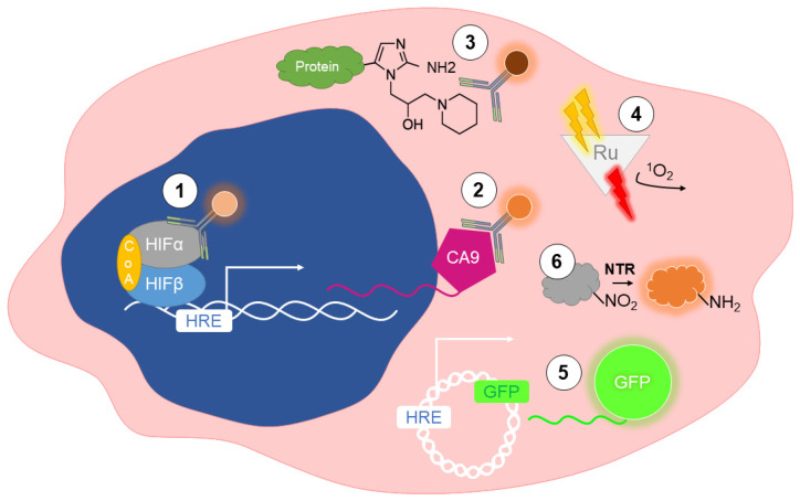
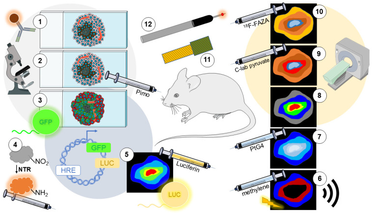
The rapid proliferation of cancer cells combined with deficient vessels cause regions of nutrient and O deprivation in solid tumors. Some cancer cells can adapt to these extreme hypoxic conditions and persist to promote cancer progression. Intratumoral hypoxia has been consistently associated with a worse patient prognosis. In vitro, 3D models of spheroids or organoids can recapitulate spontaneous O gradients in solid tumors. Likewise, in vivo murine models of cancer reproduce the physiological levels of hypoxia that have been measured in human tumors. Given the potential clinical importance of hypoxia in cancer progression, there is an increasing need to design methods to measure O concentrations. O levels can be directly measured with needle-type probes, both optical and electrochemical. Alternatively, indirect, noninvasive approaches have been optimized, and include immunolabeling endogenous or exogenous markers. Fluorescent, phosphorescent, and luminescent reporters have also been employed experimentally to provide dynamic measurements of O in live cells or tumors. In medical imaging, modalities such as MRI and PET are often the method of choice. This review provides a comparative overview of the main methods utilized to detect hypoxia in cell culture and preclinical models of cancer.
癌细胞的快速增殖加上血管不足,导致实体瘤中的营养和氧气供应不足。一些癌细胞可以适应这些极端缺氧的环境,并持续存在以促进癌症的进展。肿瘤内缺氧与患者预后较差密切相关。在体外,球体或类器官的 3D 模型可以重现实体瘤中自发的氧气梯度。同样,在体内癌症小鼠模型中重现了在人类肿瘤中测量到的生理水平的缺氧。鉴于缺氧在癌症进展中的潜在临床重要性,越来越需要设计方法来测量氧气浓度。氧气水平可以使用针型探头直接测量,包括光学和电化学方法。或者,已经优化了间接、非侵入性的方法,包括免疫标记内源性或外源性标记物。荧光、磷光和发光报告基因也被实验性地用于提供活细胞或肿瘤中氧气的动态测量。在医学成像中,MRI 和 PET 等方式通常是首选方法。本文综述了用于检测细胞培养和癌症临床前模型中缺氧的主要方法。