Friedrich Tiemo, Schalla Martha Anna, Goebel-Stengel Miriam, Kobelt Peter, Rose Matthias, Stengel Andreas
Charité Center for Internal Medicine and Dermatology, Department for Psychosomatic Medicine, Charite-Universitätsmedizin Berlin, Corporate Member of Freie Universität Berlin, Humboldt-Universität zu Berlin and Berlin Institute of Health, 12203 Berlin, Germany.
Department of Internal Medicine, Helios Kliniken GmbH, 78628 Rottweil, Germany.
Brain Sci. 2022 Jan 20;12(2):135. doi: 10.3390/brainsci12020135.
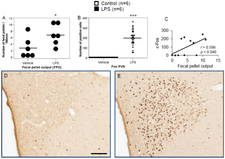
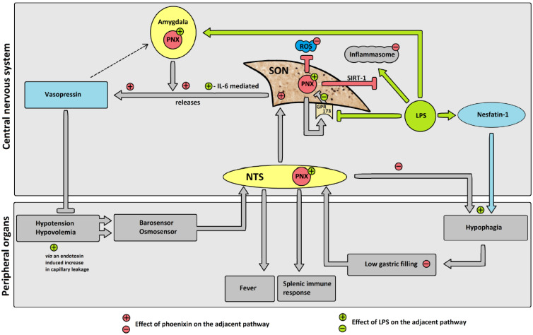
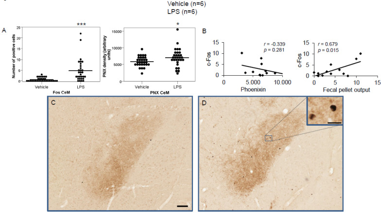
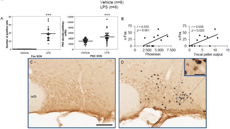
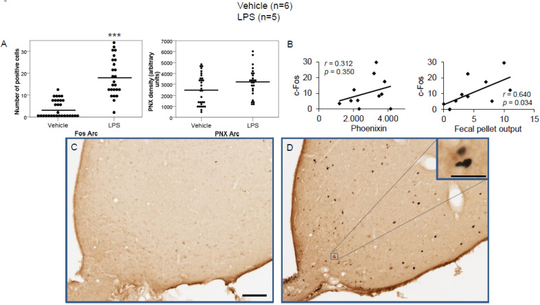
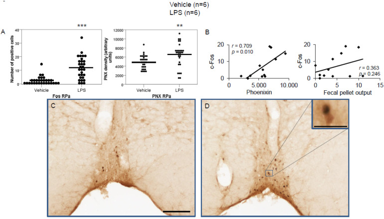
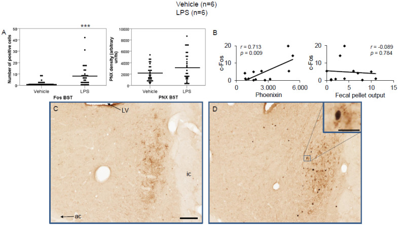
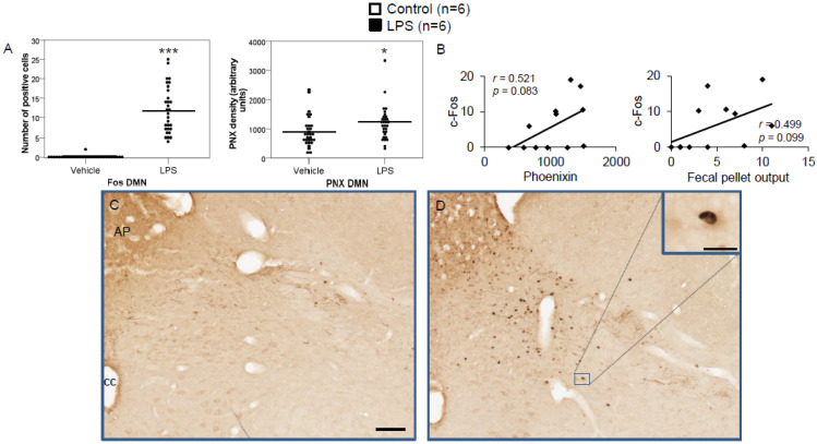
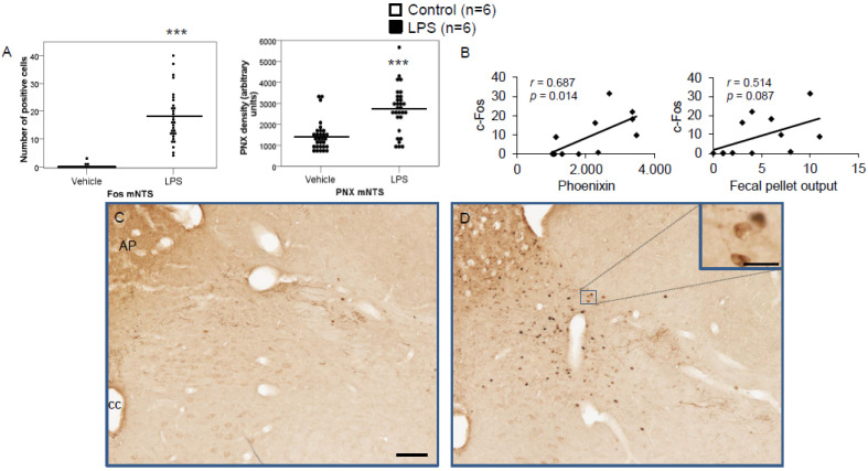
Due to phoenixin's role in restraint stress and glucocorticoid stress, as well as its recently shown effects on the inflammasome, we aimed to investigate the effects of lipopolysaccharide (LPS)-induced inflammatory stress on the activity of brain nuclei-expressing phoenixin. Male Sprague Dawley rats ( = 6/group) were intraperitoneally injected with either LPS or control (saline). Brains were processed for c-Fos and phoenixin immunohistochemistry and the resulting slides were evaluated using ImageJ software. c-Fos was counted and phoenixin was evaluated using densitometry. LPS stress significantly increased c-Fos expression in the central amygdaloid nucleus (CeM, 7.2-fold), supraoptic nucleus (SON, 34.8 ± 17.3 vs. 0.0 ± 0.0), arcuate nucleus (Arc, 4.9-fold), raphe pallidus (RPa, 5.1-fold), bed nucleus of the stria terminalis (BSt, 5.9-fold), dorsal motor nucleus of the vagus nerve (DMN, 89-fold), and medial part of the nucleus of the solitary tract (mNTS, 121-fold) compared to the control-injected group ( < 0.05). Phoenixin expression also significantly increased in the CeM (1.2-fold), SON (1.5-fold), RPa (1.3-fold), DMN (1.3-fold), and mNTS (1.9-fold, < 0.05), leading to a positive correlation between c-Fos and phoenixin in the RPa, BSt, and mNTS ( < 0.05). In conclusion, LPS stress induces a significant increase in activity in phoenixin immunoreactive brain nuclei that is distinctively different from restraint stress.
由于凤凰蛋白在束缚应激和糖皮质激素应激中的作用,以及其最近显示出的对炎性小体的影响,我们旨在研究脂多糖(LPS)诱导的炎性应激对表达凤凰蛋白的脑核活性的影响。雄性Sprague Dawley大鼠(每组 = 6只)腹腔注射LPS或对照(生理盐水)。对大脑进行c-Fos和凤凰蛋白免疫组织化学处理,并使用ImageJ软件对所得玻片进行评估。计数c-Fos,并使用光密度测定法评估凤凰蛋白。与注射对照的组相比,LPS应激显著增加了中央杏仁核(CeM,7.2倍)、视上核(SON,34.8 ± 17.3对0.0 ± 0.0)、弓状核(Arc,4.9倍)、中缝苍白核(RPa,5.1倍)、终纹床核(BSt,5.9倍)、迷走神经背运动核(DMN,89倍)和孤束核内侧部(mNTS,121倍)中c-Fos的表达(P < 0.05)。CeM(1.2倍)、SON(1.5倍)、RPa(1.3倍)、DMN(1.3倍)和mNTS(1.9倍,P < 0.05)中凤凰蛋白的表达也显著增加,导致RPa、BSt和mNTS中c-Fos与凤凰蛋白之间呈正相关(P < 0.05)。总之,LPS应激诱导凤凰蛋白免疫反应性脑核的活性显著增加,这与束缚应激明显不同。