• 文献检索
  • 文档翻译
  • 深度研究
  • 学术资讯
  • Suppr Zotero 插件Zotero 插件
  • 邀请有礼
  • 套餐&价格
  • 历史记录
应用&插件
Suppr Zotero 插件Zotero 插件浏览器插件Mac 客户端Windows 客户端微信小程序
定价
高级版会员购买积分包购买API积分包
服务
文献检索文档翻译深度研究API 文档MCP 服务
关于我们
关于 Suppr公司介绍联系我们用户协议隐私条款
关注我们

Suppr 超能文献

核心技术专利:CN118964589B侵权必究
粤ICP备2023148730 号-1Suppr @ 2026

文献检索

告别复杂PubMed语法,用中文像聊天一样搜索,搜遍4000万医学文献。AI智能推荐,让科研检索更轻松。

立即免费搜索

文件翻译

保留排版,准确专业,支持PDF/Word/PPT等文件格式,支持 12+语言互译。

免费翻译文档

深度研究

AI帮你快速写综述,25分钟生成高质量综述,智能提取关键信息,辅助科研写作。

立即免费体验

使用人工智能驱动的安卓应用程序从胸部计算机断层扫描中检测新型冠状病毒肺炎。

Detecting COVID-19 from chest computed tomography scans using AI-driven android application.

作者信息

Verma Aryan, Amin Sagar B, Naeem Muhammad, Saha Monjoy

机构信息

Department of Computer Science and Engineering, National Institute of Technology, Hamirpur, HP, 177005, India.

Department of Radiology and Imaging Sciences, Emory University School of Medicine, Atlanta, GA, 30322, USA.

出版信息

Comput Biol Med. 2022 Apr;143:105298. doi: 10.1016/j.compbiomed.2022.105298. Epub 2022 Feb 20.

DOI:10.1016/j.compbiomed.2022.105298
PMID:35220076
原文链接:https://pmc.ncbi.nlm.nih.gov/articles/PMC8858433/
Abstract

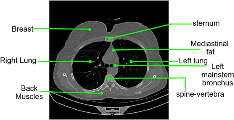
The COVID-19 (coronavirus disease 2019) pandemic affected more than 186 million people with over 4 million deaths worldwide by June 2021. The magnitude of which has strained global healthcare systems. Chest Computed Tomography (CT) scans have a potential role in the diagnosis and prognostication of COVID-19. Designing a diagnostic system, which is cost-efficient and convenient to operate on resource-constrained devices like mobile phones would enhance the clinical usage of chest CT scans and provide swift, mobile, and accessible diagnostic capabilities. This work proposes developing a novel Android application that detects COVID-19 infection from chest CT scans using a highly efficient and accurate deep learning algorithm. It further creates an attention heatmap, augmented on the segmented lung parenchyma region in the chest CT scans which shows the regions of infection in the lungs through an algorithm developed as a part of this work, and verified through radiologists. We propose a novel selection approach combined with multi-threading for a faster generation of heatmaps on a Mobile Device, which reduces the processing time by about 93%. The neural network trained to detect COVID-19 in this work is tested with a F1 score and accuracy, both of 99.58% and sensitivity of 99.69%, which is better than most of the results in the domain of COVID diagnosis from CT scans. This work will be beneficial in high-volume practices and help doctors triage patients for the early diagnosis of COVID-19 quickly and efficiently.

摘要

到2021年6月,新冠疫情(2019冠状病毒病)已影响全球超过1.86亿人,导致400多万人死亡。其规模给全球医疗系统带来了压力。胸部计算机断层扫描(CT)在新冠诊断和预后评估中具有潜在作用。设计一种经济高效且便于在手机等资源受限设备上操作的诊断系统,将提高胸部CT扫描的临床应用,并提供快速、移动且可及的诊断能力。这项工作提出开发一种新型安卓应用程序,该程序使用高效准确的深度学习算法从胸部CT扫描中检测新冠感染。它还会创建一个注意力热图,叠加在胸部CT扫描中分割出的肺实质区域上,通过作为这项工作一部分开发并经放射科医生验证的算法显示肺部的感染区域。我们提出一种结合多线程的新颖选择方法,以便在移动设备上更快地生成热图,这将处理时间减少了约93%。在这项工作中训练用于检测新冠的神经网络,F1分数和准确率均为99.58%,灵敏度为99.69%,优于CT扫描新冠诊断领域的大多数结果。这项工作将有利于大规模实践,并帮助医生快速有效地对患者进行分诊,以便早期诊断新冠。

https://cdn.ncbi.nlm.nih.gov/pmc/blobs/75bd/8858433/28bd0af17619/gr19_lrg.jpg
https://cdn.ncbi.nlm.nih.gov/pmc/blobs/75bd/8858433/94c76258764f/ga1_lrg.jpg
https://cdn.ncbi.nlm.nih.gov/pmc/blobs/75bd/8858433/3f4ee1878944/gr1_lrg.jpg
https://cdn.ncbi.nlm.nih.gov/pmc/blobs/75bd/8858433/b4cf5776258f/gr2_lrg.jpg
https://cdn.ncbi.nlm.nih.gov/pmc/blobs/75bd/8858433/04f8a521a1e3/gr3_lrg.jpg
https://cdn.ncbi.nlm.nih.gov/pmc/blobs/75bd/8858433/f12813cfacb0/gr4_lrg.jpg
https://cdn.ncbi.nlm.nih.gov/pmc/blobs/75bd/8858433/a5d18ade7b8b/gr5_lrg.jpg
https://cdn.ncbi.nlm.nih.gov/pmc/blobs/75bd/8858433/712c2503b4b7/gr6_lrg.jpg
https://cdn.ncbi.nlm.nih.gov/pmc/blobs/75bd/8858433/0f28551d78fd/gr7_lrg.jpg
https://cdn.ncbi.nlm.nih.gov/pmc/blobs/75bd/8858433/ec628e851c11/gr8_lrg.jpg
https://cdn.ncbi.nlm.nih.gov/pmc/blobs/75bd/8858433/df4a336dd16b/gr9_lrg.jpg
https://cdn.ncbi.nlm.nih.gov/pmc/blobs/75bd/8858433/9f11e8bee988/gr10_lrg.jpg
https://cdn.ncbi.nlm.nih.gov/pmc/blobs/75bd/8858433/0f83b91391cf/gr11_lrg.jpg
https://cdn.ncbi.nlm.nih.gov/pmc/blobs/75bd/8858433/00db180971a6/gr12_lrg.jpg
https://cdn.ncbi.nlm.nih.gov/pmc/blobs/75bd/8858433/0c50eb2bf6d9/gr13_lrg.jpg
https://cdn.ncbi.nlm.nih.gov/pmc/blobs/75bd/8858433/28cccedbf252/gr14_lrg.jpg
https://cdn.ncbi.nlm.nih.gov/pmc/blobs/75bd/8858433/f1ef9b0cd289/gr15_lrg.jpg
https://cdn.ncbi.nlm.nih.gov/pmc/blobs/75bd/8858433/7698622d4988/gr16_lrg.jpg
https://cdn.ncbi.nlm.nih.gov/pmc/blobs/75bd/8858433/cca667c6cd15/gr17_lrg.jpg
https://cdn.ncbi.nlm.nih.gov/pmc/blobs/75bd/8858433/97d6b5c1299b/gr18_lrg.jpg
https://cdn.ncbi.nlm.nih.gov/pmc/blobs/75bd/8858433/28bd0af17619/gr19_lrg.jpg

相似文献

1
Detecting COVID-19 from chest computed tomography scans using AI-driven android application.使用人工智能驱动的安卓应用程序从胸部计算机断层扫描中检测新型冠状病毒肺炎。
Comput Biol Med. 2022 Apr;143:105298. doi: 10.1016/j.compbiomed.2022.105298. Epub 2022 Feb 20.
2
Computed tomography-based COVID-19 triage through a deep neural network using mask-weighted global average pooling.基于计算机断层扫描的 COVID-19 分诊的深度神经网络方法,使用口罩加权全局平均池化。
Front Cell Infect Microbiol. 2023 Mar 3;13:1116285. doi: 10.3389/fcimb.2023.1116285. eCollection 2023.
3
From community-acquired pneumonia to COVID-19: a deep learning-based method for quantitative analysis of COVID-19 on thick-section CT scans.从社区获得性肺炎到 COVID-19:一种基于深度学习的 CT 厚层扫描 COVID-19 定量分析方法。
Eur Radiol. 2020 Dec;30(12):6828-6837. doi: 10.1007/s00330-020-07042-x. Epub 2020 Jul 18.
4
Deep learning-based triage and analysis of lesion burden for COVID-19: a retrospective study with external validation.基于深度学习的 COVID-19 病变负担分类和分析:一项具有外部验证的回顾性研究。
Lancet Digit Health. 2020 Oct;2(10):e506-e515. doi: 10.1016/S2589-7500(20)30199-0. Epub 2020 Sep 22.
5
A semi-supervised learning approach for COVID-19 detection from chest CT scans.一种用于从胸部CT扫描中检测新冠肺炎的半监督学习方法。
Neurocomputing (Amst). 2022 Sep 7;503:314-324. doi: 10.1016/j.neucom.2022.06.076. Epub 2022 Jun 23.
6
A deep learning-based application for COVID-19 diagnosis on CT: The Imaging COVID-19 AI initiative.基于深度学习的 CT 新冠肺炎诊断应用:影像 COVID-19 AI 计划。
PLoS One. 2023 May 2;18(5):e0285121. doi: 10.1371/journal.pone.0285121. eCollection 2023.
7
An automated diagnosis and classification of COVID-19 from chest CT images using a transfer learning-based convolutional neural network.利用基于迁移学习的卷积神经网络对 chest CT 图像进行 COVID-19 的自动诊断和分类。
Comput Biol Med. 2022 May;144:105383. doi: 10.1016/j.compbiomed.2022.105383. Epub 2022 Mar 10.
8
COVID-DSNet: A novel deep convolutional neural network for detection of coronavirus (SARS-CoV-2) cases from CT and Chest X-Ray images.COVID-DSNet:一种新型深度卷积神经网络,用于从 CT 和胸部 X 光图像中检测冠状病毒(SARS-CoV-2)病例。
Artif Intell Med. 2022 Dec;134:102427. doi: 10.1016/j.artmed.2022.102427. Epub 2022 Oct 17.
9
Artificial intelligence for assistance of radiology residents in chest CT evaluation for COVID-19 pneumonia: a comparative diagnostic accuracy study.人工智能辅助放射科住院医师对 COVID-19 肺炎进行胸部 CT 评估的诊断准确性比较研究。
Acta Radiol. 2023 Jun;64(6):2104-2110. doi: 10.1177/02841851231162085. Epub 2023 Mar 8.
10
A wavelet-based deep learning pipeline for efficient COVID-19 diagnosis via CT slices.一种基于小波的深度学习管道,用于通过CT切片高效诊断新冠肺炎。
Appl Soft Comput. 2022 Oct;128:109401. doi: 10.1016/j.asoc.2022.109401. Epub 2022 Jul 29.

引用本文的文献

1
Application of comprehensive geriatric assessment in oncology nursing: A literature review on optimizing treatment decisions and patient outcomes.综合老年评估在肿瘤护理中的应用:关于优化治疗决策和患者结局的文献综述
World J Clin Oncol. 2025 Apr 24;16(4):104785. doi: 10.5306/wjco.v16.i4.104785.
2
COVID-19 detection in lung CT slices using Brownian-butterfly-algorithm optimized lightweight deep features.使用布朗尼蝴蝶算法优化的轻量级深度特征在肺部CT切片中检测COVID-19
Heliyon. 2024 Mar 2;10(5):e27509. doi: 10.1016/j.heliyon.2024.e27509. eCollection 2024 Mar 15.
3
Mobile Apps for COVID-19 Detection and Diagnosis for Future Pandemic Control: Multidimensional Systematic Review.
移动应用程序在新冠病毒检测和诊断方面的应用,为未来的大流行控制提供了支持:多维系统评价。
JMIR Mhealth Uhealth. 2024 Feb 22;12:e44406. doi: 10.2196/44406.
4
A Systematic Review on Deep Structured Learning for COVID-19 Screening Using Chest CT from 2020 to 2022.2020年至2022年基于胸部CT的COVID-19筛查深度结构化学习系统综述
Healthcare (Basel). 2023 Aug 24;11(17):2388. doi: 10.3390/healthcare11172388.
5
Generative Adversarial Network (Generative Artificial Intelligence) in Pediatric Radiology: A Systematic Review.生成对抗网络(生成式人工智能)在儿科放射学中的应用:一项系统综述。
Children (Basel). 2023 Aug 10;10(8):1372. doi: 10.3390/children10081372.
6
Deep-Learning-Based COVID-19 Diagnosis and Implementation in Embedded Edge-Computing Device.基于深度学习的新冠病毒疾病诊断及在嵌入式边缘计算设备中的实现
Diagnostics (Basel). 2023 Apr 3;13(7):1329. doi: 10.3390/diagnostics13071329.
7
Institutional Strategies to Maintain and Grow Imaging Research During the COVID-19 Pandemic.机构策略以维持和促进 COVID-19 大流行期间的影像研究。
Acad Radiol. 2023 Apr;30(4):631-639. doi: 10.1016/j.acra.2022.12.045. Epub 2023 Jan 6.
8
An Artificial Intelligence-as-a-Service Architecture for deep learning model embodiment on low-cost devices: A case study of COVID-19 diagnosis.一种用于在低成本设备上实现深度学习模型的人工智能即服务架构:以新冠病毒疾病诊断为例
Appl Soft Comput. 2023 Feb;134:110014. doi: 10.1016/j.asoc.2023.110014. Epub 2023 Jan 13.
9
DBOF-COVIDNet: A Framework of Deep Bayesian Optimization and Fusion-Assisted Optimal Deep Features for COVID-19 Classification Using Chest X-ray and MRI Scans.DBOF-COVIDNet:一种用于使用胸部X光和MRI扫描进行COVID-19分类的深度贝叶斯优化与融合辅助最优深度特征框架。
Diagnostics (Basel). 2022 Dec 29;13(1):101. doi: 10.3390/diagnostics13010101.
10
Combating Covid-19 using machine learning and deep learning: Applications, challenges, and future perspectives.利用机器学习和深度学习抗击新冠疫情:应用、挑战及未来展望
Array (N Y). 2023 Mar;17:100271. doi: 10.1016/j.array.2022.100271. Epub 2022 Dec 10.