Redondo-Camós María, Cattaneo Gabriele, Perellón-Alfonso Ruben, Alviarez-Schulze Vanessa, Morris Timothy P, Solana-Sanchez Javier, España-Irla Goretti, Delgado-Gallén Selma, Pachón-García Catherine, Albu Sergiu, Zetterberg Henrik, Tormos Josep M, Pascual-Leone Alvaro, Bartres-Faz David
Institut Guttmann, Institut Universitari de Neurorehabilitació adscrit a la Universitat Autónoma de Barcelona, Barcelona, Spain.
Departament de Medicina, Facultat de Medicina, Universitat Autònoma de Barcelona, Barcelona, Spain.
Front Psychol. 2022 Feb 10;13:813444. doi: 10.3389/fpsyg.2022.813444. eCollection 2022.
The prefrontal cortex (PFC) plays a crucial role in cognition, particularly in executive functions. Cortical reactivity measured with Transcranial Magnetic Stimulation combined with Electroencephalography (TMS-EEG) is altered in pathological conditions, and it may also be a marker of cognitive status in middle-aged adults. In this study, we investigated the associations between cognitive measures and TMS evoked EEG reactivity and explored whether the effects of this relationship were related to neurofilament light chain levels (NfL), a marker of neuroaxonal damage.
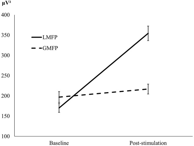
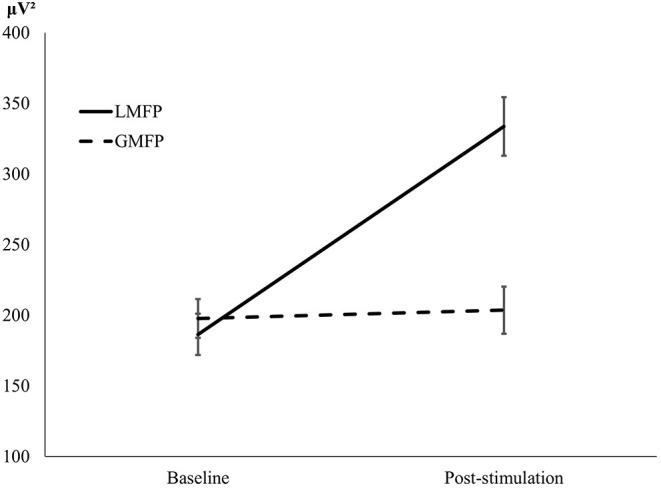
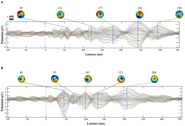
Fifty two healthy middle-aged adults (41-65 years) from the Barcelona Brain Health Initiative cohort underwent TMS-EEG, a comprehensive neuropsychological assessment, and a blood test for NfL levels. Global and Local Mean-Field Power (GMFP/LMFP), two measures of cortical reactivity, were quantified after left prefrontal cortex (L-PFC) stimulation, and cognition was set as the outcome of the regression analysis. The left inferior parietal lobe (L-IPL) was used as a control stimulation condition.
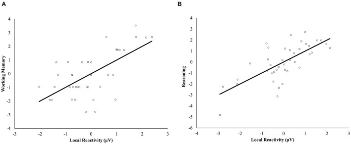
Local reactivity was significantly associated with working memory and reasoning only after L-PFC stimulation. No associations were found between NfL and cognition. These specific associations were independent of the status of neuroaxonal damage indexed by the NfL biomarker and remained after adjusting for age, biological sex, and education.
Our results demonstrate that TMS evoked EEG reactivity at the L-PFC, but not the L-IPL, is related to the cognitive status of middle-aged individuals and independent of NfL levels, and may become a valuable biomarker of frontal lobe-associated cognitive function.
前额叶皮质(PFC)在认知中起着关键作用,尤其是在执行功能方面。经颅磁刺激结合脑电图(TMS-EEG)测量的皮质反应性在病理状态下会发生改变,它也可能是中年成年人认知状态的一个标志物。在本研究中,我们调查了认知测量与TMS诱发的脑电图反应性之间的关联,并探讨了这种关系的影响是否与神经丝轻链水平(NfL)有关,NfL是神经轴突损伤的一个标志物。
来自巴塞罗那脑健康倡议队列的52名健康中年成年人(41 - 65岁)接受了TMS-EEG、全面的神经心理学评估以及NfL水平的血液检测。在左侧前额叶皮质(L-PFC)刺激后,对全局和局部平均场功率(GMFP/LMFP)这两种皮质反应性测量指标进行量化,并将认知设定为回归分析的结果。将左侧顶下小叶(L-IPL)用作对照刺激条件。
仅在L-PFC刺激后,局部反应性与工作记忆和推理显著相关。未发现NfL与认知之间存在关联。这些特定关联独立于由NfL生物标志物所索引的神经轴突损伤状态,并且在调整年龄、生物学性别和教育程度后仍然存在。
我们的结果表明,L-PFC而非L-IPL处的TMS诱发的脑电图反应性与中年个体的认知状态相关且独立于NfL水平,并且可能成为额叶相关认知功能的一个有价值的生物标志物。