Saber Manal Mohamed
Clinical Pathology Department, Faculty of Medicine, Minia University, Minia 61519, Egypt.
Antibodies (Basel). 2022 Feb 16;11(1):15. doi: 10.3390/antib11010015.
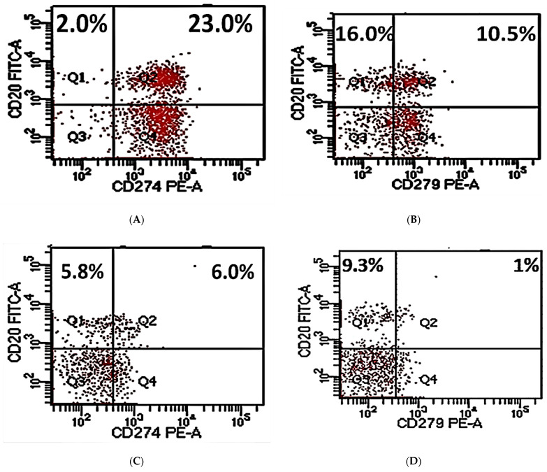
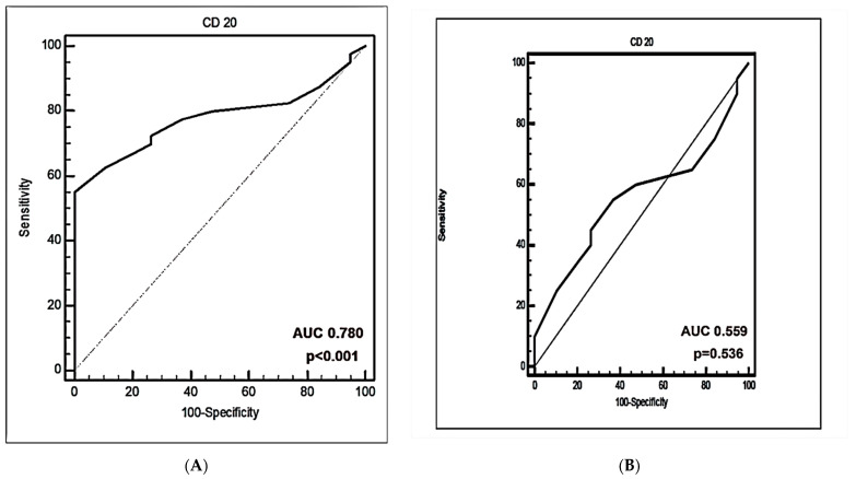
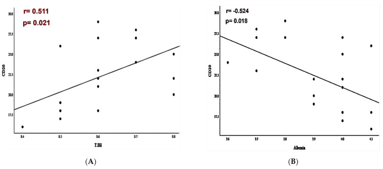
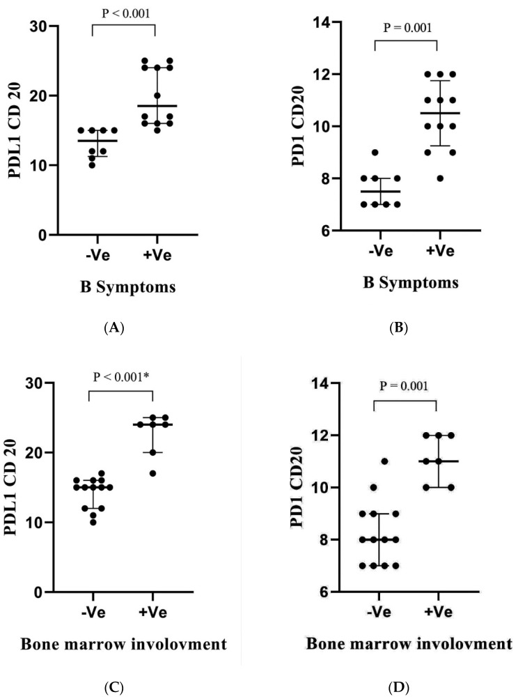
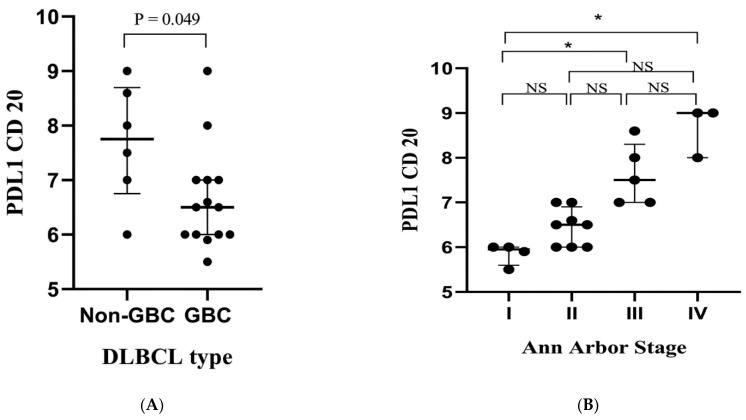
This study aimed to investigate PD-L1 and PD-1 expression in circulating CD20+ cells in diffuse larger B-cell lymphoma (DLBCL) and to evaluate the predictive and diagnostic performance of PD-L1 versus PD-1 expression in circulating CD20+ cells in DLBCL. Percentages of CD20+, PD-L1+CD20+, and PD-1+CD20+ cells were measured by flow cytometry in 40 DLBCL blood samples and 19 healthy controls. The DLBCL patient group was subdivided into 20 newly diagnosed patients with no treatment yet and 20 patients that had finished six cycles of CHOP therapy. Percentages of PD-L1+CD20+ and PD-1+CD20+ cells were highly significantly increased in pre-therapy patients in comparison to healthy volunteers ( < 0.001). Meanwhile, a significant decrease in percentages of PD-L1+CD20+ and PD-1+CD20+ was observed in post-CHOP therapy patients in comparison to pre-therapy patients ( < 0.001). PD-L1+CD20+ cells were significantly decreased in post-therapy patients when compared to normal controls ( < 0.001), while not for PD-1+CD20+ cells. A strong significant positive correlation between percentages of PD-L1+CD20+ and PD-1+CD20+ was detected in DLBCL patients ( < 0.001). In the pre-therapy group, high PD-L1+CD20+ and PD-1+CD20+ percentages were correlated with serum LDH levels ( = 0.021, < 0.001). High percentages of PD-1+CD20+ were found in DLBCL patients with splenomegaly ( = 0.027). The results revealed that patients with advanced tumor stages, poor ECOG performance, and non-GCB DLBCL type had increased percentages of PD-L1+CD20+ and PD-1+CD20+ cells. Moreover, PD-L1+CD20+ % and PD-1+CD20+ % were significantly increased in DLBCL patients with bone marrow involvement or B symptoms. The superiority of PD-L1+CD20+ over PD-1+CD20+ was more profound in DLBCL prediction [AUC: 1.0] and in discriminating newly diagnosed patients [AUC: 1.0]. The findings suggest that increased PD-L1/PD-1 expression in peripheral CD20 cells may serve as a companion diagnostic marker for DLBCL. Moreover, percentages of PD-L1+CD20+ cells have better diagnostic performance with higher sensitivity and specificity than PD-1+CD20+ %.
本研究旨在调查弥漫性大B细胞淋巴瘤(DLBCL)循环CD20+细胞中PD-L1和PD-1的表达情况,并评估PD-L1与PD-1在DLBCL循环CD20+细胞中表达的预测和诊断性能。通过流式细胞术检测了40例DLBCL血样和19例健康对照中CD20+、PD-L1+CD20+和PD-1+CD20+细胞的百分比。DLBCL患者组分为20例尚未接受治疗的新诊断患者和20例已完成六个周期CHOP治疗的患者。与健康志愿者相比,治疗前患者中PD-L1+CD20+和PD-1+CD20+细胞的百分比显著升高(<0.001)。同时,与治疗前患者相比,CHOP治疗后患者中PD-L1+CD20+和PD-1+CD20+的百分比显著降低(<0.001)。与正常对照相比,治疗后患者中PD-L1+CD20+细胞显著减少(<0.001),而PD-1+CD20+细胞则不然。在DLBCL患者中检测到PD-L1+CD20+和PD-1+CD20+百分比之间存在强显著正相关(<0.001)。在治疗前组中,高PD-L1+CD20+和PD-1+CD20+百分比与血清LDH水平相关(=0.021,<0.001)。在有脾肿大的DLBCL患者中发现高百分比的PD-1+CD20+(=0.027)。结果显示,肿瘤分期晚、ECOG体能状态差和非生发中心B细胞型DLBCL患者的PD-L1+CD20+和PD-1+CD20+细胞百分比增加。此外,在有骨髓受累或B症状的DLBCL患者中,PD-L1+CD20+%和PD-1+CD20+%显著增加。在DLBCL预测中[AUC:1.0]以及在鉴别新诊断患者中[AUC:1.0],PD-L1+CD20+比PD-1+CD20+更具优势。这些发现表明,外周CD20细胞中PD-L1/PD-1表达增加可能作为DLBCL的伴随诊断标志物。此外,PD-L1+CD20+细胞百分比比PD-1+CD20+%具有更好的诊断性能,敏感性和特异性更高。